Cuticle itching represents a surprisingly common dermatological concern that affects millions of individuals worldwide, yet remains poorly understood by both patients and healthcare providers. The delicate periungual tissues surrounding your nails serve as critical protective barriers, making them particularly susceptible to irritation, infection, and inflammatory responses that manifest as persistent pruritus. Understanding the complex interplay between anatomical structures, pathophysiological mechanisms, and environmental factors becomes essential for addressing this uncomfortable condition effectively.
The sensation of itchy cuticles often signals underlying disruption in the nail unit’s homeostasis, ranging from simple dryness to complex autoimmune conditions. Modern lifestyle factors , including frequent hand sanitiser use, chemical exposure, and aggressive manicuring practices, have contributed to an increased prevalence of cuticle-related symptoms. This comprehensive examination explores the multifaceted nature of cuticle pruritus, providing insights into both immediate relief strategies and long-term management approaches.
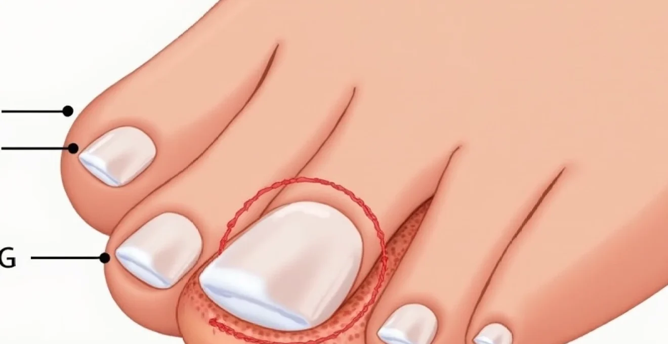
Dermatological anatomy of the cuticle and nail matrix
The nail unit comprises a sophisticated arrangement of specialised tissues, each contributing to nail formation and protection. Understanding this complex anatomy proves crucial for comprehending why cuticles become itchy and how various treatments target specific structures. The nail apparatus extends far beyond the visible nail plate, encompassing proximal and lateral nail folds, the nail matrix, nail bed, and hyponychium.
Eponychium structure and keratinocyte production
The eponychium, commonly referred to as the cuticle, consists of a thin layer of keratinised tissue that extends from the proximal nail fold onto the nail plate surface. This structure contains specialised keratinocytes that produce protective proteins and lipids, forming a crucial barrier against pathogen invasion. Disruption of keratinocyte function through chemical exposure or mechanical trauma frequently triggers inflammatory cascades that manifest as itching sensations.
Keratinocyte turnover in the eponychium occurs approximately every 14-21 days, significantly faster than other skin areas. This rapid cellular renewal makes the tissue particularly vulnerable to nutritional deficiencies, hormonal fluctuations, and environmental stressors. When keratinocyte production becomes dysregulated, the resulting structural abnormalities often present as thickened, irregular cuticles accompanied by persistent pruritus.
Hyponychium barrier function and moisture retention
The hyponychium forms the seal between the nail plate and nail bed at the fingertip, preventing bacterial invasion and maintaining optimal moisture levels. This tissue contains numerous sebaceous glands and sweat ducts that contribute to the nail unit’s hydrolipidic film. Compromised hyponychium function leads to increased transepidermal water loss, creating the dry, irritated conditions that frequently trigger itching episodes.
Moisture retention within the hyponychium depends on intact lipid barrier function and adequate hydration. Environmental factors such as low humidity, excessive washing, or chemical exposure can disrupt this delicate balance. Barrier dysfunction often presents with visible signs including peeling skin, micro-fissures, and the characteristic tight, itchy sensation many patients describe.
Perionychium inflammation patterns and sensory nerve distribution
The perionychium encompasses all soft tissue surrounding the nail plate, containing a rich network of sensory nerve endings that detect mechanical, thermal, and chemical stimuli. These nerve fibres, primarily derived from the digital nerves, show remarkable sensitivity to inflammatory mediators released during immune responses. Understanding this neurological component explains why minor inflammation can produce disproportionately intense itching sensations.
Inflammatory patterns within the perionychium follow predictable pathways, typically beginning with vasodilation and increased vascular permeability. Sensory nerve activation occurs through direct mechanical pressure from tissue swelling and chemical stimulation by inflammatory mediators. This dual mechanism explains why anti-inflammatory treatments often provide rapid relief from cuticle itching, even before visible improvements occur.
Nail fold vascularisation and immune response mechanisms
The proximal and lateral nail folds contain an extensive capillary network that facilitates rapid immune cell recruitment during inflammatory responses. This vascularisation pattern makes the nail folds particularly reactive to systemic conditions affecting circulation or immune function. Vascular changes often precede visible inflammation, explaining why patients may experience itching before other symptoms develop.
Immune surveillance within the nail folds involves resident dendritic cells, macrophages, and lymphocytes that monitor for foreign antigens and tissue damage. When these cells detect threats, they initiate cascading immune responses that can persist long after the initial trigger resolves. This prolonged activation frequently manifests as chronic cuticle itching that resists conventional topical treatments.
Pathophysiological mechanisms triggering cuticle pruritus
The sensation of itching, or pruritus, results from complex interactions between sensory neurons, inflammatory mediators, and central nervous system processing. In cuticle tissues, these mechanisms become particularly pronounced due to the high density of nerve endings and the tissue’s exposure to environmental irritants. Understanding these pathways enables targeted therapeutic interventions that address root causes rather than simply masking symptoms.
Histamine release from mast cell degranulation
Mast cells within periungual tissues serve as primary mediators of allergic and inflammatory responses, releasing histamine and other bioactive compounds when activated by specific triggers. These triggers include mechanical trauma from aggressive manicuring, chemical allergens in nail products, and bacterial toxins from infected cuticles. Histamine release produces immediate vasodilation, increased vascular permeability, and direct stimulation of pruriceptive nerve fibres.
The magnitude of mast cell degranulation varies significantly between individuals, explaining why identical exposures produce different symptom intensities. Genetic polymorphisms affecting histamine metabolism and receptor sensitivity contribute to this variability. Chronic mast cell activation can lead to tissue remodelling and persistent hypersensitivity, creating a cycle of ongoing symptoms that requires comprehensive management approaches.
Prostaglandin E2 and inflammatory mediator cascade
Prostaglandin E2 (PGE2) functions as a key inflammatory mediator in cuticle tissues, amplifying pain and itch sensations while promoting tissue swelling and redness. PGE2 synthesis increases dramatically following tissue injury, infection, or chemical irritation. This compound not only directly stimulates sensory neurons but also enhances their sensitivity to other inflammatory mediators, creating a synergistic effect that intensifies symptoms.
The inflammatory mediator cascade involves multiple cytokines, including interleukin-1β, tumour necrosis factor-α, and various chemokines that attract immune cells to the affected area. Cascade amplification explains why minor cuticle irritation can progress to significant inflammation if left untreated. Interrupting this cascade early through appropriate interventions prevents symptom escalation and tissue damage.
Neuropeptide substance P and CGRP involvement
Substance P and calcitonin gene-related peptide (CGRP) represent crucial neuropeptides involved in itch sensation transmission and neurogenic inflammation. These compounds are released from sensory nerve terminals in response to various stimuli, including mechanical pressure, heat, and chemical irritants. Their release creates a positive feedback loop, as both peptides enhance their own synthesis and release while promoting local inflammation.
Neurogenic inflammation mediated by substance P and CGRP contributes significantly to chronic cuticle itching conditions. These neuropeptides increase vascular permeability, stimulate immune cell activation, and sensitise surrounding nerve fibres to subsequent stimuli. Neuropeptide modulation through topical treatments or systemic medications offers promising therapeutic approaches for persistent symptoms.
Transient receptor potential channel activation
Transient receptor potential (TRP) channels function as molecular sensors for various environmental stimuli, including temperature changes, chemical irritants, and mechanical pressure. Several TRP channel subtypes are highly expressed in cuticle tissues, particularly TRPV1 and TRPA1 channels that respond to capsaicin-like compounds and environmental irritants respectively. Activation of these channels triggers immediate itch sensations and inflammatory responses.
TRP channel sensitivity can become enhanced through repeated exposure to irritants or during inflammatory conditions, leading to heightened reactivity to normally innocuous stimuli. This sensitisation process explains why individuals with chronic cuticle problems often develop reactions to previously tolerated products. Understanding TRP channel pharmacology opens new therapeutic possibilities, including topical modulators that can reset channel sensitivity to normal levels.
Infectious aetiologies and microbial colonisation
Microbial infections represent a significant cause of cuticle itching, with various pathogens capable of colonising the warm, moist environment around nail tissues. The unique anatomy of the nail unit creates microenvironments that favour different types of microorganisms, from bacteria to fungi and yeasts. Understanding these infectious processes proves essential for appropriate treatment selection and prevention of recurrent episodes.
Candida albicans overgrowth in periungual tissue
Candida albicans commonly colonises periungual tissues, particularly in individuals with compromised immune function, diabetes, or frequent water exposure. This opportunistic yeast produces enzymes that digest keratin and other proteins, creating inflammatory byproducts that trigger intense itching sensations. Candidal overgrowth often presents with characteristic white patches, tissue maceration, and persistent burning or itching symptoms that worsen with moisture exposure.
Risk factors for candidal cuticle infections include immunosuppression, antibiotic use, pregnancy, and occupational exposures to water or chemicals. The infection can spread rapidly across nail folds and may involve multiple digits simultaneously. Early recognition and treatment prevent chronic colonisation that becomes increasingly difficult to eradicate over time.
Staphylococcus aureus biofilm formation
Staphylococcus aureus represents the most common bacterial pathogen affecting cuticle tissues, capable of forming protective biofilms that resist standard antibiotic treatments. These biofilms create chronic inflammatory conditions characterised by persistent redness, swelling, and intense itching. The bacteria produce various toxins and enzymes that directly damage tissue and trigger robust immune responses.
Biofilm-associated infections often require prolonged treatment courses combining systemic antibiotics with topical antimicrobials. Biofilm disruption through mechanical debridement or specialised treatments may be necessary for complete eradication. Prevention focuses on maintaining intact cuticle barriers and avoiding trauma that provides bacterial entry points.
Pseudomonas aeruginosa nail bed invasion
Pseudomonas aeruginosa infections typically occur following nail trauma or aggressive manicuring practices that compromise tissue integrity. This opportunistic pathogen produces distinctive blue-green pigments and proteolytic enzymes that cause significant tissue damage. The infection often presents with characteristic discoloration, foul odour, and severe itching that may progress to pain if left untreated.
Pseudomonas infections require prompt recognition and aggressive treatment, as the organism can rapidly spread to deeper tissues. The pathogen shows inherent resistance to many common antibiotics, necessitating culture-guided therapy for optimal outcomes. Environmental sources include contaminated water systems, inadequately sterilised nail equipment, and artificial nail applications performed under non-sterile conditions.
Dermatophyte species and tinea unguium presentation
Dermatophyte fungi, including Trichophyton rubrum and Trichophyton mentagrophytes, commonly affect nail units and surrounding tissues. These organisms possess specialised enzymes that digest keratin, allowing invasion of nail plates and adjacent skin structures. Dermatophyte infections typically begin in cuticle areas before spreading to involve nail plates, producing characteristic thickening, discoloration, and persistent itching symptoms.
Diagnosis requires microscopic examination and fungal culture, as clinical presentation alone cannot distinguish between different species or rule out bacterial co-infections. Treatment often involves prolonged courses of antifungal medications, both topical and systemic, depending on infection extent and patient factors. Prevention strategies focus on maintaining dry conditions and avoiding contaminated environments such as public pools or shared footwear.
The complex interplay between host immunity, environmental factors, and microbial virulence determines infection outcomes and treatment requirements for cuticle-related conditions.
Contact dermatitis and chemical irritant exposure
Chemical irritants represent one of the most frequent causes of cuticle itching in modern society, with exposure occurring through occupational activities, household products, and personal care items. The thin, permeable nature of cuticle tissues makes them particularly vulnerable to chemical penetration and subsequent inflammatory responses. Understanding common irritants and their mechanisms of action enables effective prevention and treatment strategies.
Irritant contact dermatitis develops through direct toxic effects on skin cells, while allergic contact dermatitis involves immune-mediated hypersensitivity reactions to specific chemicals. Both conditions can produce intense itching, though the timing and characteristics of symptoms differ. Irritant reactions typically occur within hours of exposure and affect all exposed individuals given sufficient concentration and duration. Allergic reactions require prior sensitisation and may develop days after contact, affecting only susceptible individuals.
Common chemical irritants include formaldehyde and formaldehyde-releasing preservatives found in nail hardeners, acrylates and methacrylates in artificial nail systems, toluene sulfonamide in nail polishes, and quaternary ammonium compounds in nail treatment products. Occupational exposures include cleaning chemicals, industrial solvents, metalworking fluids, and agricultural chemicals. Even seemingly benign products like hand sanitisers can cause cuticle irritation with frequent use due to their alcohol content and preservative systems.
Prevention strategies focus on identifying and avoiding problematic chemicals through careful product selection and protective measures during exposure. Patch testing can identify specific allergens in cases of suspected allergic contact dermatitis. Treatment typically involves topical corticosteroids for acute inflammation, though chronic cases may require systemic interventions. Barrier restoration through appropriate moisturisers and protective agents helps prevent recurrent episodes and promotes healing of damaged tissues.
Systemic conditions manifesting through cuticle symptoms
Numerous systemic conditions can manifest early symptoms through changes in cuticle appearance and sensations, making careful evaluation of persistent itching essential for identifying underlying health issues. The rich vascular supply and rapid cellular turnover of periungual tissues make them sensitive indicators of systemic dysfunction. Recognising these patterns enables early intervention and appropriate specialist referral when indicated.
Autoimmune conditions frequently affect nail units, with lupus erythematosus, dermatomyositis, and scleroderma producing characteristic cuticle changes accompanied by itching or burning sensations. These conditions often present with dilated capillaries visible at the proximal nail fold, irregular cuticle overgrowth, and increased sensitivity to minor trauma. Autoimmune manifestations may precede systemic symptoms by months or years, making nail examination crucial for early diagnosis.
Endocrine disorders, particularly thyroid dysfunction and diabetes mellitus, commonly affect nail health and cuticle integrity. Hypothyroidism produces slow nail growth, thick cuticles, and increased susceptibility to fungal infections, while hyperthyroidism may cause rapid, brittle nail growth with inflamed cuticles. Diabetes affects circulation and immune function, predisposing to chronic cuticle infections and delayed healing responses that manifest as persistent itching.
Nutritional deficiencies, especially biotin, zinc, and essential fatty acids, can produce cuticle changes that include excessive dryness, cracking, and associated itching symptoms. These deficiencies may result from inadequate dietary intake, malabsorption syndromes, or increased metabolic demands during illness or pregnancy. Nutritional assessment should be considered in patients with chronic cuticle problems resistant to conventional treatments.
Early recognition of systemic conditions through cuticle examination can significantly impact patient outcomes by enabling timely diagnosis and intervention before more serious complications develop.
Therapeutic interventions and clinical management protocols
Effective management of itchy cuticles requires a systematic approach that addresses both immediate symptom relief and underlying causative factors. Treatment protocols should be individualised based on symptom severity, suspected aetiology, patient preferences, and response to initial interventions. A comprehensive approach often combines topical treatments, systemic medications when indicated, and preventive measures to reduce recurrence risk.
Topical corticosteroids remain the cornerstone of acute anti-inflammatory treatment, with potency selection based on symptom severity
and anatomical location. Mid-potency corticosteroids such as triamcinolone acetonide 0.1% provide excellent anti-inflammatory effects for most cuticle conditions without significant risk of skin atrophy with short-term use. High-potency preparations may be necessary for severe inflammatory responses but require careful monitoring to prevent adverse effects.
Topical calcineurin inhibitors, including tacrolimus and pimecrolimus, offer valuable alternatives to corticosteroids for patients requiring prolonged treatment or those at risk of steroid-related complications. These medications effectively suppress local immune responses while maintaining barrier function, making them particularly suitable for chronic inflammatory conditions affecting cuticle tissues.
Antimicrobial treatments should be selected based on suspected or confirmed pathogen identification. Topical antibiotics such as mupirocin or fusidic acid effectively treat superficial bacterial infections, while systemic antibiotics may be necessary for deeper tissue involvement or immunocompromised patients. Antifungal therapies range from topical agents like ciclopirox or amorolfine for localised infections to systemic treatments including terbinafine or itraconazole for extensive or resistant cases.
Barrier repair represents a crucial component of comprehensive treatment protocols. Ceramide-containing moisturisers help restore lipid barrier function, while urea-based preparations provide both moisturising and mild keratolytic effects that improve cuticle texture. Occlusive treatments using petrolatum-based products overnight can accelerate healing in severely compromised tissues. Consistency in barrier restoration often determines long-term treatment success and prevention of symptom recurrence.
Systemic interventions may be warranted for severe cases or those associated with underlying medical conditions. Antihistamines provide symptomatic relief for histamine-mediated itching, though sedating formulations should be used cautiously. Systemic corticosteroids are reserved for extensive inflammatory conditions that fail to respond to topical treatments, with careful consideration of benefit-risk profiles given potential adverse effects.
Preventive measures form an integral part of long-term management strategies. Patient education regarding proper nail care techniques, identification of potential irritants, and recognition of early warning signs helps prevent recurrent episodes. Regular application of protective hand creams before exposure to potential irritants can significantly reduce symptom frequency and severity. Lifestyle modifications, including dietary optimisation and stress management, may provide additional benefits for patients with chronic conditions.
Successful management of itchy cuticles requires patience and persistence, as complete resolution may take several weeks to months depending on the underlying cause and individual healing capacity.
Follow-up protocols should include regular assessment of treatment response and adjustment of therapeutic approaches based on clinical outcomes. Patients experiencing persistent symptoms despite appropriate treatment require evaluation for underlying systemic conditions or consideration of specialist dermatological referral. Documentation of treatment responses helps guide future management decisions and identifies patterns that may suggest specific aetiologies requiring targeted interventions.