Loop Electrosurgical Excision Procedure (LEEP) represents one of the most effective interventions for removing precancerous cervical tissue, yet many patients experience unexpected vaginal discharge patterns during recovery. Black discharge following this electrocautery treatment often causes considerable anxiety, though it frequently represents a normal physiological response to the healing process. Understanding the distinction between expected post-procedural discharge and potentially concerning symptoms becomes crucial for patient peace of mind and appropriate medical management. The complex interplay between cauterised tissue debris, oxidised blood products, and normal cervical healing creates various discharge characteristics that require careful interpretation within the context of individual recovery patterns.
Understanding LEEP procedure and normal Post-Operative discharge patterns
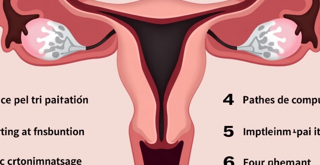
Loop electrosurgical excision procedure cervical tissue removal process
LEEP utilises a thin wire loop heated with electrical current to precisely remove abnormal cervical tissue identified through colposcopy or abnormal Pap smear results. The electrocautery process simultaneously cuts and cauterises tissue, creating a controlled zone of thermal injury that promotes haemostasis whilst removing targeted cellular abnormalities. This dual action of cutting and coagulating blood vessels minimises immediate bleeding but creates distinctive tissue changes that influence subsequent discharge patterns. The procedure typically removes a cone-shaped portion of cervical tissue, extending from the transformation zone where most precancerous changes occur.
During the electrocautery process, temperatures reaching 100-200°C create immediate tissue desiccation and protein denaturation. This thermal effect extends beyond the immediate excision site, creating a zone of coagulation necrosis that serves as a biological barrier against bleeding. However, this same thermal injury initiates a complex wound healing cascade that produces various discharge components over subsequent weeks. The application of Monsel’s paste (ferric subsulphate) following tissue removal further contributes to the chemical composition of early post-procedural discharge.
Expected vaginal discharge timeline following electrocautery treatment
Post-LEEP discharge typically evolves through predictable phases corresponding to different aspects of cervical healing. Initial discharge within the first 48 hours often contains Monsel’s paste residue, appearing dark brown or black due to iron compounds used for haemostasis. This phase may also include small amounts of charred tissue debris and oxidised blood products from the electrocautery process. The combination of these elements creates a characteristic dark, sometimes coffee-ground appearance that alarms many patients despite being entirely normal.
Between days 3-10 post-procedure, discharge patterns shift as inflammatory responses predominate. Cellular debris from healing tissues mixes with increased cervical mucus production, often creating yellow-green or brown discharge with varying consistency. The sloughing of superficial necrotic tissue during this period contributes additional dark material to vaginal discharge. By week 2-3, discharge typically transitions to clearer or white secretions as epithelial regeneration progresses and inflammatory responses subside.
Cervical healing phases and associated discharge characteristics
The inflammatory phase of cervical healing, occurring primarily during the first week post-LEEP, generates increased vascular permeability and white cell infiltration. This process produces discharge rich in inflammatory mediators, cellular debris, and protein-rich exudates. The breakdown of red blood cells within this inflammatory milieu contributes haemoglobin degradation products that may appear dark brown or black when mixed with other discharge components. Understanding these normal inflammatory responses helps patients distinguish expected healing discharge from potentially pathological symptoms.
Proliferative healing phases beginning in the second week involve rapid epithelial cell division and angiogenesis. During this period, discharge typically becomes less voluminous and lighter in colour as inflammatory processes resolve. However, intermittent dark discharge may still occur as deeper tissue layers continue healing and occasional small vessels undergo natural breakdown. The cervical transformation zone requires particularly complex healing due to the junction between squamous and glandular epithelia, potentially prolonging certain discharge patterns.
Distinguishing normal charred tissue debris from abnormal symptoms
Normal post-LEEP discharge containing charred tissue debris typically lacks offensive odour and occurs without associated systemic symptoms such as fever or significant pelvic pain. The volume of normal discharge rarely exceeds that of a typical menstrual period, and patients can usually manage symptoms with standard sanitary protection. Charred tissue debris appears as small, dark particles mixed with other discharge components, often resembling coffee grounds or pepper flakes in consistency.
Abnormal discharge patterns requiring medical evaluation include sudden increases in volume, particularly if exceeding heavy menstrual flow, or discharge accompanied by fever above 38°C. Foul-smelling discharge suggests potential bacterial overgrowth or infection, warranting prompt medical assessment. Additionally, discharge associated with severe cramping, unusual bleeding patterns, or persistent bright red bleeding beyond the first few days requires professional evaluation to exclude complications such as secondary haemorrhage or infection.
Clinical significance of black vaginal discharge after electrosurgical cervical treatment
Cauterised tissue sloughing and carbon residue elimination
Electrocautery creates a superficial layer of carbonised tissue that gradually separates from underlying healthy cervical epithelium during the healing process. This carbonised layer, or eschar, contains actual carbon particles formed during the high-temperature tissue treatment, contributing directly to black discharge components. The sloughing process typically begins 3-5 days post-procedure as inflammatory responses soften the adherence between viable and non-viable tissue layers. Carbon residue elimination continues for 1-2 weeks, with intermittent black particles appearing in discharge as deeper eschar layers separate.
The molecular structure of carbonised tissue differs significantly from normal cellular debris, creating distinctive visual and textural characteristics in vaginal discharge. These carbon particles resist normal enzymatic breakdown processes, requiring mechanical elimination through natural cervical secretions and vaginal discharge. Understanding this process helps explain why black discharge may appear intermittently rather than continuously, as eschar separation occurs in irregular fragments rather than uniform layers.
Haemoglobin oxidation and melanoidin formation in vaginal environment
Blood products trapped within cauterised tissue undergo complex chemical changes that contribute to black discharge coloration. Haemoglobin oxidation occurs rapidly in the presence of heat and oxygen, creating methemoglobin and other oxidised iron compounds with characteristic dark brown to black appearances. The acidic vaginal environment further promotes these oxidation reactions, intensifying colour changes in blood-derived discharge components. Melanoidin formation through non-enzymatic browning reactions between amino acids and reducing sugars in tissue fluids adds additional dark pigmentation to post-LEEP discharge.
These biochemical processes continue for several days following the initial procedure, explaining why black discharge may appear or intensify 3-7 days post-LEEP rather than immediately. The interaction between residual Monsel’s paste iron compounds and oxidising blood products creates particularly dark discharge that often concerns patients. However, these reactions represent normal consequences of the electrocautery process rather than pathological developments requiring intervention.
Eschar separation process and associated dark discharge components
Eschar formation following electrocautery creates a protective biological barrier over the treated cervical area, but this same barrier must eventually separate to allow normal epithelial regeneration. The separation process involves enzymatic breakdown of denatured proteins, mechanical loosening through inflammatory oedema, and gradual replacement by granulation tissue. During separation, fragments of dark, leathery eschar tissue become incorporated into vaginal discharge, creating the characteristic black or dark brown appearance that alarms many patients.
The timing of eschar separation correlates with individual healing responses, typically occurring between days 5-14 post-procedure. Some patients experience gradual, continuous separation with minimal discharge changes, whilst others have more dramatic separation events producing sudden increases in dark discharge volume.
The eschar separation process represents a critical phase of healing that, whilst producing concerning discharge changes, indicates normal progression toward complete cervical recovery.
Understanding this timeline helps patients anticipate and appropriately manage discharge changes without unnecessary anxiety.
Endocervical gland secretion changes Post-Electrocautery
Electrocautery effects extend beyond surface epithelium to influence endocervical gland function and secretion patterns. Thermal injury to glandular tissue may temporarily alter mucus production, creating thicker, more viscous secretions that trap cellular debris and blood products more effectively. These altered secretions contribute to darker discharge colours and may persist for several weeks as glandular epithelium regenerates. Endocervical gland dysfunction following LEEP may also create temporary changes in discharge pH and bacterial flora that influence overall discharge characteristics.
The complex architecture of endocervical crypts means that some glandular tissue may remain temporarily compromised even after surface healing appears complete. This can result in intermittent dark discharge episodes weeks after the initial procedure as deeper glandular structures continue healing. The restoration of normal glandular function typically requires 4-6 weeks, corresponding to complete cervical healing timelines established through clinical research.
Pathological black discharge indicators requiring medical intervention
Bacterial vaginosis and anaerobic infection warning signs
Whilst black discharge often represents normal post-LEEP healing, certain characteristics suggest pathological processes requiring immediate medical evaluation. Bacterial vaginosis developing after LEEP procedures may produce dark, foul-smelling discharge due to anaerobic bacterial overgrowth in the altered cervical environment. The disruption of normal vaginal flora following electrocautery creates opportunities for pathogenic bacterial colonisation, particularly in patients with predisposing factors such as diabetes or immunocompromisation. Anaerobic infections produce characteristic fishy or putrid odours that distinguish pathological discharge from normal healing secretions.
Additional warning signs of bacterial infection include discharge associated with burning sensations, unusual itching, or systemic symptoms such as fever and malaise. The volume of infected discharge typically exceeds normal post-procedural expectations, and the consistency may become frothy or unusually thick. Patients experiencing these symptoms require prompt antibiotic therapy to prevent progression to more serious pelvic infections that could compromise long-term cervical health and fertility.
Cervical stenosis complications with retained menstrual blood
Cervical stenosis represents a rare but serious complication of LEEP procedures that can create black discharge through blood retention mechanisms. Excessive scarring during healing may narrow the cervical os sufficiently to impair normal menstrual flow, causing blood to accumulate within the uterine cavity or cervical canal. This retained blood undergoes degradation and oxidation processes that produce characteristic black discharge when eventually expelled.
Cervical stenosis complications require prompt medical intervention to prevent serious reproductive health consequences including infection and infertility.
Patients at higher risk for cervical stenosis include those with extensive tissue removal, previous cervical procedures, or healing complications such as infection. The development of stenosis typically becomes apparent 4-8 weeks post-procedure when normal menstrual patterns should have resumed. Symptoms may include absent or significantly reduced menstrual flow, severe menstrual cramping, or intermittent expulsion of dark, old blood. Early recognition and treatment of stenosis prevent more serious complications such as haematometra or ascending infection.
Secondary haemorrhage risk factors and coagulated blood accumulation
Secondary haemorrhage following LEEP procedures typically occurs 7-14 days post-procedure as eschar separation exposes underlying blood vessels before complete epithelialisation. When bleeding occurs gradually or intermittently, blood may accumulate within the cervical canal or upper vagina, undergoing coagulation and oxidation processes that create black discharge when eventually expelled. Risk factors for secondary haemorrhage include large excision volumes, patient bleeding disorders, and premature physical activity during healing.
Coagulated blood accumulation may present as sudden episodes of dark discharge mixed with clots or tissue-like material, often accompanied by cramping or pelvic pressure. Unlike normal post-procedural discharge, secondary haemorrhage typically involves larger volumes and may include bright red bleeding intermixed with dark material. Patients experiencing sudden increases in discharge volume, particularly with associated cramping or clot passage, require immediate medical evaluation to exclude active bleeding requiring intervention.
Pelvic inflammatory disease development following LEEP
Pelvic inflammatory disease (PID) represents a serious but uncommon complication of LEEP procedures that may produce dark discharge through inflammatory and infectious processes. The temporary disruption of cervical barrier function following electrocautery creates potential pathways for ascending bacterial infection, particularly in patients with pre-existing risk factors such as multiple sexual partners or sexually transmitted infections. PID-associated discharge typically becomes increasingly purulent and malodorous over time, often progressing from initial dark coloration to frankly purulent yellow-green secretions.
Early signs of PID development include pelvic pain extending beyond normal post-procedural discomfort, fever above 38°C, and discharge changes accompanied by urinary symptoms or abnormal bleeding. The progression from dark discharge to obviously infected secretions may occur rapidly, making early recognition crucial for preventing serious reproductive health consequences. Patients with suspected PID require immediate broad-spectrum antibiotic therapy and close monitoring to prevent complications such as tubo-ovarian abscess formation or chronic pelvic pain syndromes.
Duration and resolution patterns of Post-LEEP black discharge
Normal black discharge following LEEP procedures typically resolves within 2-3 weeks as cervical healing progresses through predictable phases. The most intense period of dark discharge usually occurs during days 5-10 post-procedure, corresponding to peak eschar separation and inflammatory responses. Individual variation in healing rates means some patients may experience black discharge for up to 4 weeks, whilst others notice resolution within 10-14 days. Factors influencing discharge duration include the extent of tissue removal, individual healing responses, adherence to post-procedural restrictions, and presence of contributing medical conditions.
The gradual transition from black discharge to normal cervical secretions provides valuable indicators of healing progression. Patients typically notice decreasing discharge volume and colour intensity as healing advances, with intermittent dark episodes becoming less frequent over time. Complete resolution of abnormal discharge usually coincides with restoration of normal cervical epithelium, typically occurring 4-6 weeks post-procedure. However, some patients may experience occasional dark discharge episodes for up to 8 weeks as deeper cervical structures complete healing processes.
Persistent black discharge beyond 6-8 weeks warrants medical evaluation to exclude complications such as incomplete healing, infection, or cervical stenosis. Additionally, discharge that initially improves but then worsens or returns after apparent resolution may indicate developing complications requiring intervention.
Monitoring discharge patterns throughout the healing period provides valuable insights into recovery progress and helps identify potential complications before they become serious health threats.
Patients should maintain detailed records of discharge characteristics, including volume, colour, odour, and associated symptoms, to facilitate accurate assessment during follow-up appointments.
Evidence-based management protocols for abnormal Post-Procedural discharge
Evidence-based management of post-LEEP discharge begins with comprehensive patient education regarding normal healing patterns and warning signs requiring immediate medical attention. Clinical protocols emphasise the importance of distinguishing physiological healing responses from pathological complications through systematic symptom assessment and appropriate diagnostic testing. Conservative management approaches focus on supporting natural healing processes whilst monitoring for signs of complications that require active intervention.
When pathological discharge patterns are suspected, diagnostic approaches typically include speculum examination to assess cervical healing, collection of discharge specimens for microscopic examination and culture, and evaluation of associated symptoms such as fever or pelvic pain. Treatment protocols vary depending on identified causes, ranging from topical antimicrobial therapy for localised infections to systemic antibiotics for more extensive inflammatory processes. Some cases may require minor surgical interventions such as cervical dilation for stenosis or silver nitrate application for persistent bleeding.
Follow-up protocols generally recommend initial assessment at 2-3 weeks post-procedure to evaluate healing progress and address any discharge concerns. Patients experiencing normal healing patterns typically require only routine follow-up at 6 months for repeat cytological examination. However, those with complicated healing may need more frequent monitoring to ensure complete resolution of abnormal discharge patterns and prevention of long-term complications. Individualised management approaches consider patient risk factors, symptom severity, and response to initial interventions when determining appropriate treatment escalation.
Long-term cervical health monitoring after loop electrosurgical excision
Long-term cervical health monitoring following LEEP procedures requires systematic assessment of healing completeness, cancer surveillance through regular cytological screening, and evaluation of potential late complications such as cervical incompetence or stenosis. The relationship between initial discharge patterns and long-term outcomes remains an active area of clinical research, with some studies suggesting that prolonged abnormal discharge may predict increased risk of healing complications or cervical dysfunction.
Established surveillance protocols recommend cytological examination at 6-month intervals for the first two years following LEEP, with transition to annual screening thereafter in patients demonstrating consistently normal results. The persistence of high-risk HPV strains following tissue removal necessitates enhanced monitoring protocols, as viral persistence correlates with increased risk of recurrent cervical dysplasia or progression to invasive carcinoma. HPV co-testing alongside cytological examination provides superior sensitivity for detecting early recurrent disease compared to cytology alone, particularly in the immediate post-procedure period when inflammatory changes may mask cellular abnormalities.
Reproductive health considerations following LEEP procedures include monitoring for cervical incompetence during subsequent pregnancies, particularly in patients who required extensive tissue removal or experienced healing complications. The relationship between cervical shortening following LEEP and preterm birth risk remains controversial, with current evidence suggesting minimal impact for most patients but potential increased surveillance needs for high-risk cases. Long-term fertility outcomes generally remain excellent following appropriately performed LEEP procedures, though some patients may experience subtle changes in cervical mucus production that could theoretically impact natural conception rates.
Late complications such as cervical stenosis may develop months or years following the initial procedure, particularly in patients with extensive scarring or recurrent procedures. Regular assessment of menstrual patterns and cervical patency during routine gynecological examinations helps identify developing stenosis before symptoms become severe. Preventive interventions such as cervical dilation may be recommended for patients at high risk of stenosis development, though the optimal timing and technique for such interventions continues to evolve based on emerging clinical evidence.
The integration of discharge pattern assessment into long-term monitoring protocols provides valuable clinical information about cervical healing adequacy and potential complications. Patients who experienced prolonged or complicated discharge patterns during initial healing may benefit from more frequent follow-up examinations to ensure complete epithelialisation and normal cervical function restoration. Documentation of discharge characteristics during the acute healing phase can inform future clinical decision-making, particularly regarding the appropriateness of repeat procedures or alternative treatment approaches should abnormal cytology recur.
Comprehensive long-term monitoring following LEEP procedures requires integration of symptom assessment, cytological surveillance, and clinical examination to optimise patient outcomes whilst minimising unnecessary interventions or anxiety-provoking procedures.
Patient education regarding long-term cervical health maintenance emphasises the importance of regular screening compliance, HPV vaccination when appropriate, and lifestyle modifications that support optimal cervical health. The psychological impact of abnormal discharge experiences during initial healing may influence patient adherence to follow-up recommendations, making supportive counselling and clear communication about expected outcomes essential components of comprehensive care. Understanding the normal progression from concerning black discharge to complete healing helps patients maintain confidence in their treatment outcomes and commit to necessary long-term monitoring protocols that ensure optimal cervical health preservation throughout their reproductive years.