Discovering a hard lump in the rectal area can be an alarming experience that prompts immediate concern about potential underlying conditions. These palpable masses within or around the rectum represent a diverse spectrum of pathological processes, ranging from benign haemorrhoidal disease to more serious malignant conditions. The complexity of anorectal anatomy, combined with the rich vascular supply and lymphatic drainage of this region, creates an environment where various disease processes can manifest as firm, indurated masses.
Understanding the differential diagnosis of rectal lumps requires comprehensive knowledge of both common and rare conditions affecting the anorectal region. The clinical presentation of these masses often overlaps, making accurate diagnosis challenging without proper examination and diagnostic workup. Factors such as location, consistency, mobility, associated symptoms, and patient demographics all contribute to narrowing the differential diagnosis and guiding appropriate management strategies.
Haemorrhoids and thrombosed external haemorrhoids as primary rectal mass causes
Haemorrhoidal disease represents the most prevalent cause of palpable rectal masses, affecting approximately 50% of adults over age 50. The pathophysiology involves engorgement and prolapse of the haemorrhoidal cushions, which are normal anatomical structures composed of arteriovenous connections, smooth muscle, and connective tissue. When these cushions become pathologically enlarged due to increased venous pressure, chronic straining, or tissue degeneration, they can present as firm, palpable masses within the anal canal or protruding externally.
The haemorrhoidal plexus consists of three main cushions positioned at the left lateral, right anterior, and right posterior aspects of the anal canal. These cushions contribute to anal continence by providing up to 15% of resting anal pressure. When inflammation, thrombosis, or chronic prolapse occurs, the normally soft tissue becomes indurated and may feel like a hard lump during digital examination or self-palpation.
Grade IV prolapsed internal haemorrhoids presenting as hard rectal lumps
Grade IV internal haemorrhoids represent the most severe form of haemorrhoidal prolapse, characterised by permanently protruding tissue that cannot be manually reduced. These prolapsed haemorrhoids often become chronically inflamed, leading to fibrotic changes and induration that creates the sensation of a hard rectal lump. The chronic exposure to external environment results in squamous metaplasia of the initially columnar epithelium, further contributing to tissue hardening.
Patients with Grade IV haemorrhoids frequently report a constant feeling of rectal fullness, incomplete evacuation, and the presence of a firm mass that may cause discomfort during sitting or walking. The prolapsed tissue often becomes oedematous and may develop ulcerations, particularly when subjected to trauma from wiping or clothing friction. These complications can lead to secondary bacterial infection, further increasing tissue induration and patient discomfort.
Thrombosed external haemorrhoid pathophysiology and clinical presentation
Thrombosed external haemorrhoids develop when blood clots form within the external haemorrhoidal plexus, typically following acute increases in intra-abdominal pressure during straining, heavy lifting, or prolonged sitting. The thrombosis creates a rapidly developing, extremely painful, and characteristically hard lump at the anal margin. Unlike internal haemorrhoids, external haemorrhoids are covered by squamous epithelium and innervated by somatic nerves, making them exquisitely painful when thrombosed.
The clinical presentation typically involves sudden onset of severe anal pain accompanied by a visible, firm, bluish-purple nodule at the anal verge. The thrombosed haemorrhoid feels rock-hard to palpation and may range in size from a few millimetres to several centimetres in diameter. Patients often describe the pain as throbbing or burning, frequently worse with sitting, bowel movements, or any activity that increases pelvic pressure.
Differential diagnosis between haemorrhoidal complexes and perianal masses
Distinguishing haemorrhoidal disease from other perianal masses requires careful clinical assessment and understanding of anatomical landmarks. True haemorrhoids arise from the haemorrhoidal cushions and maintain specific positional relationships to the dentate line, whereas other masses may occur at any location around the anal circumference. Haemorrhoidal tissue typically demonstrates a characteristic grape-like appearance when prolapsed, with visible vascular patterns beneath the mucosa or skin surface.
The mobility and reducibility of haemorrhoidal masses also differ from other conditions. Early-grade internal haemorrhoids may be reducible with gentle pressure, while external haemorrhoids remain fixed at the anal margin. In contrast, perianal abscesses present as fluctuant masses with surrounding erythema and warmth, while skin tags appear as soft, fleshy protrusions without the vascular engorgement characteristic of active haemorrhoidal disease.
Chronic haemorrhoidal disease complications leading to fibrotic tissue formation
Long-standing haemorrhoidal disease frequently results in chronic inflammatory changes that lead to fibrotic tissue formation, creating persistently hard masses in the anal region. The repeated cycles of inflammation, thrombosis, and healing result in replacement of normal elastic tissue with dense fibrous connective tissue. This process transforms initially soft, reducible haemorrhoidal tissue into firm, non-reducible masses that may be mistaken for more sinister pathology.
The fibrotic changes associated with chronic haemorrhoidal disease often create irregular, nodular surfaces that can be palpated as multiple hard areas within what was previously smooth haemorrhoidal tissue. These changes may be accompanied by the development of anal skin tags, which represent stretched skin that fails to retract following resolution of acute haemorrhoidal swelling. The combination of fibrotic haemorrhoidal tissue and associated skin tags can create a complex landscape of firm masses around the anal opening.
Anorectal malignancies and precancerous lesions
Malignant conditions of the anorectal region, while less common than benign causes, represent the most serious potential explanation for hard rectal lumps. The incidence of anorectal cancers has been steadily increasing, particularly among younger demographics, making early recognition and appropriate referral crucial for optimal patient outcomes. These malignancies can arise from different tissue types within the anorectal complex, each presenting with distinct characteristics and requiring specific management approaches.
The anatomical complexity of the anorectal junction, where different epithelial types converge, creates an environment susceptible to various malignant transformations. Rectal adenocarcinomas typically arise from glandular epithelium above the dentate line , while squamous cell carcinomas more commonly develop in the anal canal and perianal region. The rich lymphatic drainage of this area facilitates early metastatic spread, emphasising the importance of prompt diagnosis and staging when malignancy is suspected.
Adenocarcinoma of the rectum presenting with palpable rectal masses
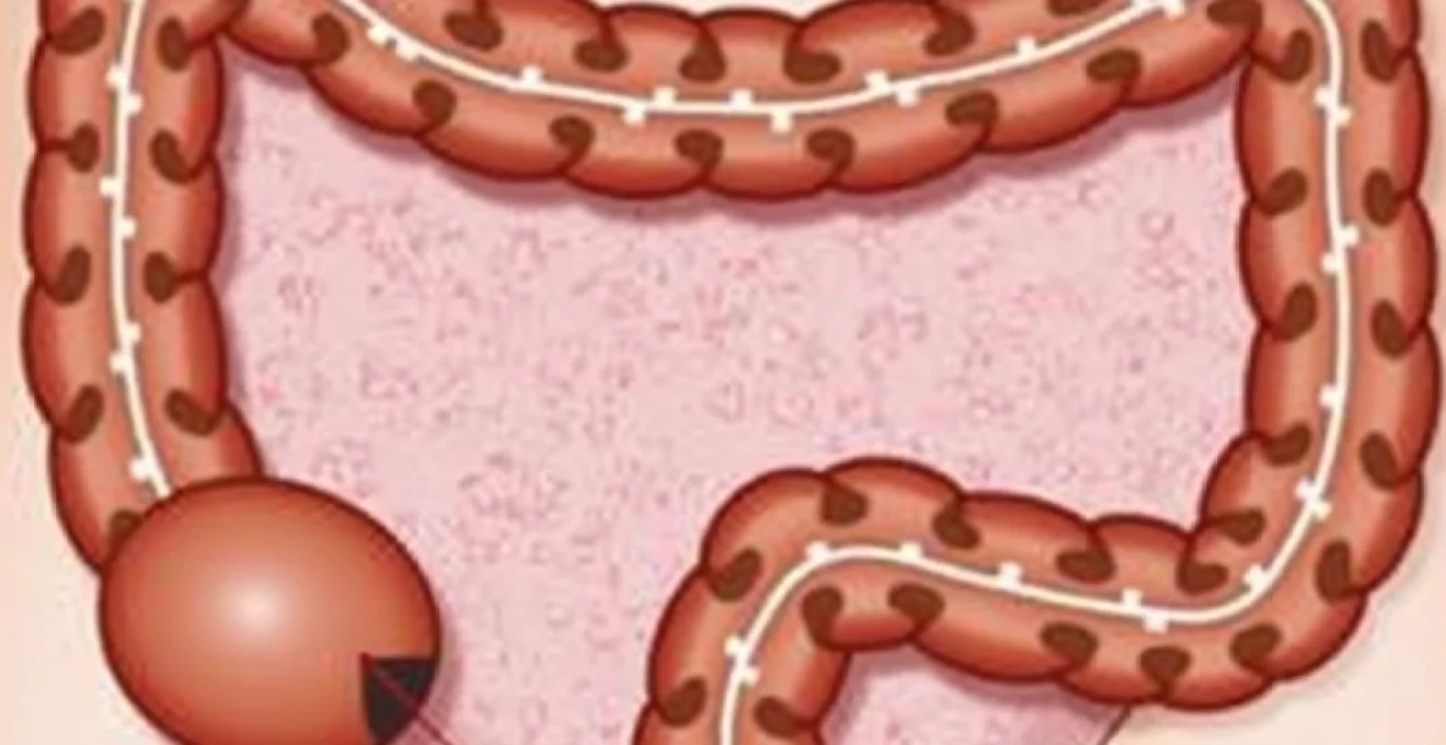
Rectal adenocarcinoma represents approximately 30% of all colorectal cancers and typically manifests as a firm, irregular mass within the rectal wall that can be palpated during digital rectal examination. These tumours most commonly develop in the distal rectum, within 15 centimetres of the anal verge, making them accessible to digital palpation in many cases. The characteristic feeling of a rectal adenocarcinoma is that of a hard, fixed, irregular mass with rolled edges and possible ulceration of the overlying mucosa.
Early-stage rectal adenocarcinomas may present as small, firm nodules within the rectal wall, while advanced tumours can create large, obstructing masses that significantly narrow the rectal lumen. The tumour consistency varies depending on histological characteristics, with well-differentiated adenocarcinomas often feeling firmer due to increased fibrous stroma, while poorly differentiated tumours may have a softer consistency but demonstrate more aggressive local invasion patterns.
Squamous cell carcinoma of the anal canal and perianal region
Anal squamous cell carcinoma has shown increasing incidence rates, particularly associated with human papillomavirus infection, and typically presents as a firm, irregular mass within the anal canal or at the anal margin. These tumours often develop from precancerous lesions such as anal intraepithelial neoplasia and may initially be mistaken for benign conditions like haemorrhoids or anal fissures. The characteristic presentation involves a hard, often ulcerated lesion that may bleed easily and cause persistent anal pain or discomfort.
The clinical distinction between anal squamous cell carcinoma and benign anal conditions can be challenging, as both may present with similar symptoms including bleeding, pain, and palpable masses. However, cancerous lesions typically demonstrate progressive growth, irregular borders, and failure to respond to conservative treatments. The induration associated with anal cancer often extends beyond the visible lesion margins, reflecting local tissue invasion and inflammatory response.
Gastrointestinal stromal tumours (GIST) in the rectoanal junction
Gastrointestinal stromal tumours affecting the rectum are rare but important causes of hard rectal masses, representing less than 5% of all rectal malignancies. These tumours arise from the interstitial cells of Cajal and typically present as firm, well-circumscribed masses within the rectal wall. GIST tumours often grow to considerable size before becoming symptomatic , and their location in the rectum may make them palpable as hard, round masses during digital examination.
The clinical behaviour of rectal GIST differs from adenocarcinomas, with these tumours often growing as exophytic masses that may cause mechanical symptoms such as tenesmus, altered bowel habits, or a sensation of incomplete evacuation. The firm consistency reflects the spindle cell morphology typical of GIST, and these tumours may achieve significant dimensions while remaining relatively asymptomatic due to their submucosal location and expansile rather than infiltrative growth pattern.
High-grade dysplastic polyps and villous adenomas with malignant potential
Large villous adenomas and polyps containing high-grade dysplasia can present as firm masses within the rectum, particularly when they develop fibrotic changes or undergo partial malignant transformation. These lesions represent important precancerous conditions that require careful evaluation and management due to their significant risk of harboring invasive carcinoma. Villous adenomas typically feel softer than invasive carcinomas but may develop areas of induration corresponding to regions of high-grade dysplasia or early invasive changes.
The clinical challenge with large adenomatous polyps lies in determining the presence and extent of malignant transformation through clinical examination alone. While benign adenomatous tissue typically feels soft and mobile, areas of induration within these lesions may indicate malignant change and require urgent histopathological evaluation. The sessile nature of many villous adenomas makes them feel like broad-based, firm areas within the rectal wall rather than discrete, mobile masses.
Infectious and inflammatory conditions creating rectal induration
Infectious and inflammatory processes affecting the anorectal region frequently result in tissue induration and mass formation through various pathophysiological mechanisms. These conditions can create diagnostic challenges due to their ability to mimic malignant processes, particularly when chronic inflammation leads to fibrotic tissue changes. The rich vascular supply and lymphatic drainage of the anorectal region, combined with its exposure to bacterial flora, creates an environment conducive to various infectious processes that may result in palpable masses.
Understanding the temporal evolution of infectious and inflammatory masses is crucial for accurate diagnosis and appropriate management. Acute infectious processes typically present with rapid onset of symptoms and associated systemic features , while chronic inflammatory conditions may develop insidiously over months or years, creating firm masses that can be difficult to distinguish from malignant lesions without appropriate diagnostic workup.
Perianal abscesses and chronic Fistula-in-Ano tract formation
Perianal abscesses represent acute infectious processes that typically begin in the anal glands and can progress to form palpable masses in various anatomical spaces around the anorectal region. The initial presentation usually involves a rapidly developing, extremely painful, fluctuant mass accompanied by systemic signs of infection including fever, malaise, and leukocytosis. However, partially treated or chronic abscesses may develop thick fibrous walls that create firm, indurated masses that persist even after drainage attempts.
Chronic fistula-in-ano tracts develop in approximately 30-50% of patients following perianal abscess drainage and can create palpable induration along their course through the perirectal tissues. These tracts often become surrounded by fibrous tissue and chronic inflammatory infiltrate, creating linear or nodular areas of induration that can be traced from the internal opening at the dentate line to the external opening on the perianal skin. Complex fistula tracts may create multiple areas of induration corresponding to secondary tracks and extensions.
Crohn’s disease perianal manifestations and stricturing complications
Crohn’s disease affects the perianal region in approximately 80% of patients with colonic involvement and can create various types of hard masses through different pathophysiological mechanisms. The transmural inflammation characteristic of Crohn’s disease leads to fibrotic stricture formation, chronic fistulous disease, and abscess formation, all of which may present as palpable rectal masses. Perianal Crohn’s disease often creates complex anatomical distortion with multiple interconnected inflammatory masses and fibrotic tissue replacement.
The chronic nature of Crohn’s disease inflammation results in progressive fibrotic changes that can create firm, irregular masses within the rectal wall and perianal tissues. These masses may be associated with anal stenosis, making digital examination difficult and potentially painful for patients. The combination of chronic inflammation, fibrosis, and associated complications such as fistulas and abscesses creates a complex clinical picture that requires multidisciplinary management approaches.
Actinomycosis and chronic granulomatous infections of the rectum
Actinomycosis represents a rare but important chronic bacterial infection that can affect the anorectal region and create firm, indurated masses that may be mistaken for malignancy. This condition typically develops following mucosal injury and involves anaerobic, gram-positive bacteria that create characteristic sulfur granules within infected tissues. The chronic inflammatory response results in extensive fibrosis and induration that can create large, firm masses extending throughout the perirectal tissues.
Other chronic granulomatous infections, including tuberculosis and atypical mycobacterial infections, may also create hard rectal masses through similar mechanisms of chronic inflammation and fibrotic tissue replacement. These conditions typically develop insidiously over months and may be associated with systemic symptoms such as weight loss, fever, and night sweats. The indurated masses associated with chronic granulomatous infections often demonstrate characteristic histopathological features that distinguish them from malignant processes.
Post-radiation proctitis fibrotic changes and rectal wall thickening
Radiation proctitis following pelvic radiotherapy for prostate, cervical, or rectal cancers can result in chronic inflammatory changes leading to significant rectal wall induration and mass formation. The pathophysiology involves radiation-induced vascular injury, chronic ischaemia, and progressive fibrotic replacement of normal rectal wall architecture. These changes typically develop 6-24 months following radiation exposure and may continue to progress for years after treatment completion.
The chronic fibrotic changes associated with radiation proctitis can create diffuse rectal wall thickening that feels like a firm, irregular mass during digital examination. The induration may be accompanied by mucosal ulceration, bleeding, and stricture formation that can significantly impact bowel function. Distinguishing radiation-induced fibrosis from recurrent malignancy represents a significant clinical challenge that often requires advanced imaging and tissue sampling for definitive diagnosis.
Benign rectal masses and structural abnormalities
Various benign conditions can create palpable masses within the rectum, ranging from congenital abnormalities to acquired structural changes. These conditions often present diagnostic challenges due to their potential to mimic more serious pathology, particularly when they become indurated through chronic inflammation or fibrotic changes. Understanding the characteristic features of benign rectal masses is essential for appropriate clinical management and patient reassurance while ensuring that more serious conditions are not overlooked.
Rectal polyps represent one of the most common benign causes of palpable rectal masses, with hyperplastic polyps and adenomatous polyps demonstrating different consistency and malignant potential. Large pedunculated polyps may feel like mobile, firm masses within the rectal lumen, while
sessile polyps may create more diffuse areas of induration within the rectal wall. The clinical significance of these lesions depends largely on their histological characteristics, size, and location within the rectum.
Rectal lipomas represent another category of benign masses that can occasionally become palpable, particularly when they achieve significant size or develop inflammatory changes. These fatty tumours typically feel soft and mobile but may become firmer when subjected to chronic irritation or when they develop a fibrous capsule. Submucosal lipomas can create the sensation of a smooth, round mass within the rectal wall that moves freely with manipulation.
Endometriosis affecting the rectovaginal septum can create firm nodular masses that may be palpable through the rectal wall, particularly in women of reproductive age. These masses typically demonstrate cyclical changes corresponding to menstrual cycles and may become more prominent and tender during menstruation. The fibrotic reaction surrounding endometriotic implants often creates a characteristic firm, irregular texture that can be distinguished from other types of rectal masses through careful clinical correlation with patient symptoms and timing.
Systemic conditions manifesting as rectal induration
Various systemic conditions can manifest with rectal induration and mass formation as part of their broader clinical presentation. These conditions often involve inflammatory, infiltrative, or metabolic processes that affect multiple organ systems, with the anorectal region being one site of involvement. Recognition of these systemic conditions requires careful attention to associated clinical features and laboratory abnormalities that may provide clues to the underlying diagnosis.
Amyloidosis represents a rare but important systemic condition that can involve the rectum, creating firm, waxy masses within the rectal wall. Primary systemic amyloidosis may present with rectal involvement as an early manifestation, while secondary amyloidosis typically occurs in the context of chronic inflammatory conditions such as inflammatory bowel disease or chronic infections. The amyloid deposits create characteristic firm, non-tender masses that may be associated with bleeding due to vascular fragility.
Lymphomatous involvement of the anorectal region, while uncommon, can create firm masses that may be mistaken for other conditions. Primary anorectal lymphomas are rare, but secondary involvement may occur in patients with systemic lymphoma, particularly in immunocompromised individuals. These masses typically feel rubbery and may be associated with systemic symptoms such as night sweats, weight loss, and lymphadenopathy in other regions.
Sarcoidosis occasionally involves the anorectal region, creating granulomatous inflammatory masses that can feel firm and irregular. The diagnosis typically requires correlation with pulmonary involvement, skin lesions, and characteristic laboratory findings including elevated serum angiotensin-converting enzyme levels. Rectal sarcoidosis may present as multiple firm nodules within the rectal wall that can be mistaken for malignant involvement without proper histopathological evaluation.
Clinical assessment and diagnostic approaches for rectal mass evaluation
The clinical evaluation of patients presenting with hard rectal lumps requires a systematic approach that combines thorough history taking, careful physical examination, and appropriate diagnostic testing. The initial assessment should focus on characterising the mass through digital rectal examination, determining associated symptoms, and identifying risk factors that may suggest specific diagnoses. The temporal evolution of symptoms, associated pain patterns, and relationship to bowel movements provide crucial diagnostic clues.
Digital rectal examination remains the cornerstone of initial assessment, allowing evaluation of mass characteristics including size, consistency, mobility, surface texture, and relationship to surrounding structures. The examination should assess the entire circumference of the rectum within reach, noting the position relative to anatomical landmarks such as the dentate line and anal verge. Proper technique involves gentle, systematic palpation using adequate lubrication and patient positioning to ensure comfort and accuracy of findings.
Anoscopy and flexible sigmoidoscopy provide direct visualisation of the rectal mucosa and allow assessment of masses that may not be readily palpable. These procedures enable tissue sampling for histopathological diagnosis when indicated and provide important information about the extent of disease involvement. The endoscopic appearance of masses can provide diagnostic clues, with malignant lesions typically demonstrating irregular surfaces, easy bleeding, and friable texture compared to benign conditions.
Advanced imaging studies including magnetic resonance imaging and computed tomography play crucial roles in staging suspected malignancies and evaluating complex inflammatory conditions. MRI provides excellent soft tissue contrast and is particularly valuable for assessing the relationship of rectal masses to surrounding structures, sphincter involvement, and lymph node status. High-resolution MRI can distinguish between different tissue types and help characterise the nature of rectal wall thickening or mass lesions.
Laboratory investigations should be tailored to the clinical presentation but may include complete blood count, inflammatory markers, tumour markers such as carcinoembryonic antigen, and specific tests for inflammatory conditions when clinically indicated. Microbiological studies including cultures and molecular testing may be appropriate when infectious aetiologies are suspected, particularly in immunocompromised patients or those with systemic symptoms.
The timing of referral to specialist services depends on clinical findings and degree of suspicion for serious pathology. Urgent referral is indicated for masses suspicious for malignancy, particularly when associated with constitutional symptoms, significant bleeding, or rapid growth. The two-week rule for suspected colorectal cancer referrals provides a framework for ensuring timely evaluation of high-risk presentations while avoiding unnecessary anxiety for patients with benign conditions.
Patient education and reassurance play important roles in managing the anxiety associated with discovering rectal masses. Clear explanation of the diagnostic process, timeline for results, and range of potential diagnoses helps patients cope with uncertainty while emphasising the importance of completing recommended evaluations. The majority of rectal masses prove to be benign conditions that can be effectively managed with appropriate treatment.