Ingrown hairs in the intimate area between the thighs represent one of the most challenging dermatological concerns for both patients and healthcare professionals. This delicate region combines unique anatomical features, increased friction, and heightened sensitivity that create the perfect storm for follicular complications. The warm, humid environment of the inner thigh area, coupled with frequent hair removal practices and tight clothing, establishes conditions where hair follicles become impacted, leading to painful inflammation and potential secondary infections.
Unlike ingrown hairs on other body areas, those occurring between the thighs require specialised treatment approaches that account for the region’s physiological complexities. The interplay between sebaceous gland activity, bacterial colonisation, and mechanical friction creates a multifaceted problem that demands comprehensive understanding and targeted intervention strategies.
Understanding folliculitis and pseudofolliculitis barbae in the inner thigh region
The pathophysiology of ingrown hairs between the thighs involves complex interactions between hair follicle architecture, skin barrier function, and environmental factors. Pseudofolliculitis barbae , whilst traditionally associated with facial hair in men, presents similarly in the inner thigh region where coarse, curly hair growth patterns predominate. The condition manifests when newly growing hair shafts curve back into the skin, creating inflammatory papules and pustules that can progress to chronic scarring if left untreated.
The inflammatory cascade begins when keratin plugs obstruct follicular openings, trapping emerging hair shafts beneath the skin surface. This mechanical obstruction triggers an immune response characterised by neutrophil infiltration and subsequent tissue damage. The resulting inflammation can persist for weeks, creating painful nodules that significantly impact quality of life and intimate comfort.
Anatomical factors contributing to hair follicle impaction
The inner thigh region possesses unique anatomical characteristics that predispose individuals to follicular complications. The convergence of multiple skin folds creates areas where moisture accumulates, promoting bacterial proliferation and softening the stratum corneum. Additionally, the hair follicles in this region typically exhibit a curved trajectory, making them more susceptible to ingrowth following hair removal procedures.
The subcutaneous adipose tissue distribution in the inner thigh creates variable skin tension patterns that can alter follicular orientation during leg movement. This dynamic environment means that hair emerging from follicles may encounter resistance from surrounding tissues, increasing the likelihood of follicular re-entry and subsequent inflammation.
Sebaceous gland dysfunction and keratin plug formation
Sebaceous glands in the inner thigh region demonstrate heightened activity due to hormonal influences and mechanical stimulation from clothing friction. This increased sebum production, when combined with desquamated keratinocytes, creates viscous plugs that obstruct follicular openings. The resulting comedone-like lesions provide an anaerobic environment conducive to bacterial proliferation.
The hyperkeratinisation process accelerates in response to chronic friction, leading to thickened follicular walls that further impede normal hair emergence. This creates a self-perpetuating cycle where mechanical irritation promotes increased keratin production, which in turn exacerbates follicular obstruction and ingrown hair formation.
Friction-induced inflammatory response mechanisms
Mechanical friction from clothing, particularly tight-fitting garments, initiates a cascade of inflammatory mediators that compromise follicular integrity. The repetitive rubbing motion damages the follicular epithelium, creating microscopic wounds that serve as entry points for opportunistic bacteria. This friction-induced trauma also stimulates the release of pro-inflammatory cytokines, perpetuating the inflammatory response long after the initial mechanical insult.
The inflammatory response involves activation of the complement system and recruitment of inflammatory cells, including mast cells, which release histamine and other vasoactive substances. This creates the characteristic erythema, oedema, and pruritus associated with ingrown hairs in the inner thigh region.
Bacterial colonisation by staphylococcus epidermidis
The warm, moist environment between the thighs provides optimal conditions for bacterial colonisation, particularly by Staphylococcus epidermidis and other coagulase-negative staphylococci. These commensal organisms can become pathogenic when follicular barriers are compromised, leading to secondary bacterial folliculitis that complicates the clinical presentation of ingrown hairs.
Biofilm formation by these bacteria creates a protective matrix that enhances antibiotic resistance and prolongs the inflammatory process. The bacterial metabolites also contribute to follicular damage by producing enzymes that degrade the follicular wall structure, facilitating deeper tissue penetration and chronic infection.
Clinical assessment and differential diagnosis protocols
Accurate clinical assessment of ingrown hairs between the thighs requires systematic evaluation to differentiate from other inflammatory skin conditions. The diagnostic process begins with comprehensive history-taking, focusing on hair removal practices, clothing preferences, and symptom timeline. Physical examination should assess lesion morphology, distribution patterns, and signs of secondary infection or scarring.
The clinical presentation typically includes erythematous papules, pustules, and occasionally larger nodules in areas corresponding to hair distribution patterns. Patients often report cyclical symptoms that correlate with hair removal activities, with peak inflammation occurring 24-48 hours post-epilation. The presence of visible trapped hairs within lesions provides pathognomonic evidence of follicular impaction.
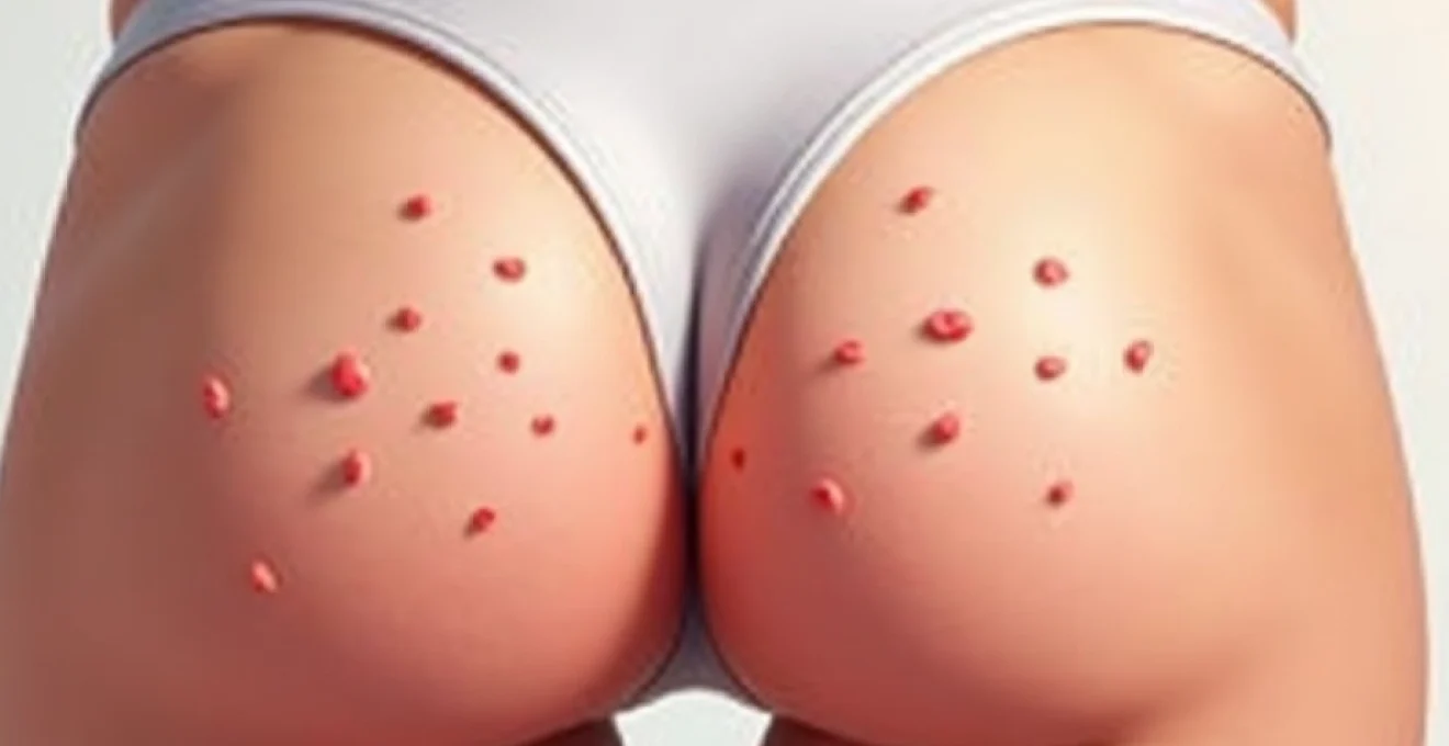
Visual identification of papular and pustular lesions
Ingrown hairs present as inflammatory papules ranging from 1-3mm in diameter, often surrounded by an erythematous halo. The lesions may progress to pustular stages, containing purulent material that reflects secondary bacterial infection. Careful inspection may reveal the characteristic “comma-shaped” or looped hair visible beneath the skin surface, particularly when using magnification techniques.
Chronic lesions may develop into firm, hyperpigmented nodules with associated scarring. These post-inflammatory changes are particularly problematic in patients with darker skin types, where hyperpigmentation can persist for months following resolution of the acute inflammatory phase.
Distinguishing hidradenitis suppurativa from ingrown hair cysts
Hidradenitis suppurativa represents a significant differential diagnosis that requires careful consideration in patients presenting with recurrent inflammatory lesions in the inner thigh region. Unlike ingrown hairs, hidradenitis suppurativa typically presents with deeper, more painful nodules that may progress to abscess formation and sinus tract development. The presence of comedone-like lesions and the characteristic “tombstone” appearance of double-ended comedones suggests hidradenitis suppurativa rather than simple follicular impaction.
The distribution pattern also differs, with hidradenitis suppurativa showing predilection for apocrine gland-rich areas and demonstrating bilateral symmetry. Additionally, hidradenitis suppurativa lesions tend to recur in the same anatomical locations, creating characteristic scarring patterns that distinguish them from the more randomly distributed ingrown hair lesions.
Dermoscopy examination techniques for follicular assessment
Dermoscopic examination enhances diagnostic accuracy by revealing subtle follicular changes not visible to the naked eye. The presence of curved or looped hairs, black dots representing fractured hair shafts, and follicular hyperkeratosis provides definitive evidence of ingrown hair pathology. Dermoscopy also enables assessment of surrounding inflammation and identification of bacterial colonisation patterns.
The technique involves systematic examination using 10-20x magnification, focusing on individual lesions and surrounding follicular structures. Polarised light dermoscopy can reveal subsurface hair structures and help differentiate ingrown hairs from other follicular disorders such as keratosis pilaris or follicular hyperkeratosis.
Fitzpatrick skin type classification impact on treatment selection
Patient skin phototype significantly influences both the clinical presentation and treatment approach for ingrown hairs. Individuals with Fitzpatrick skin types IV-VI demonstrate increased propensity for post-inflammatory hyperpigmentation, requiring modified treatment protocols that minimise risk of pigmentary changes. Additionally, these patients may show more prominent inflammatory responses to follicular impaction, necessitating earlier and more aggressive intervention.
The hair characteristics also vary with ethnicity, with patients of African descent demonstrating curved hair follicles and tightly coiled hair shafts that predispose to ingrown hair formation. These anatomical differences require tailored prevention and treatment strategies that account for the unique follicular architecture.
Topical pharmaceutical interventions and active compounds
Topical pharmaceutical management forms the cornerstone of ingrown hair treatment, utilising active compounds that target multiple aspects of the pathophysiological process. The therapeutic approach combines anti-inflammatory agents, keratolytics, and antimicrobial compounds to address follicular obstruction, reduce inflammation, and prevent secondary bacterial infection. Selection of appropriate topical agents depends on lesion severity, skin sensitivity, and individual patient factors including skin phototype and concurrent medications.
Retinoid compounds, particularly tretinoin and adapalene, demonstrate significant efficacy in normalising follicular keratinisation and reducing comedone formation. These agents work by modulating cellular differentiation and reducing follicular hyperkeratosis that contributes to hair entrapment. Clinical studies indicate that consistent retinoid application can reduce ingrown hair incidence by up to 60% when used as part of a comprehensive treatment protocol. The anti-inflammatory properties of retinoids also help reduce the inflammatory response associated with follicular impaction.
Alpha-hydroxy acids, including glycolic acid and lactic acid, provide chemical exfoliation that helps release trapped hairs and prevents future follicular obstruction. These compounds work by dissolving the intercellular cement that holds corneocytes together, facilitating natural desquamation and reducing keratin plug formation. Concentration selection typically ranges from 5-20%, with higher concentrations reserved for treatment-resistant cases under professional supervision. The pH optimisation of these formulations ensures maximum efficacy whilst minimising irritation in the sensitive inner thigh region.
Topical antimicrobial agents play a crucial role in managing secondary bacterial infection and preventing progression to more severe folliculitis. Clindamycin phosphate 1% demonstrates excellent penetration into pilosebaceous units and maintains broad-spectrum activity against gram-positive bacteria commonly implicated in follicular infections. Benzoyl peroxide, whilst primarily recognised for acne treatment, provides both antimicrobial and comedolytic activity that benefits ingrown hair management. The combination of clindamycin and benzoyl peroxide in fixed-dose formulations prevents bacterial resistance development whilst maximising therapeutic efficacy.
Professional extraction techniques and sterile procedures
Professional extraction represents a critical intervention for established ingrown hairs that fail to resolve with topical therapy alone. These procedures require meticulous attention to aseptic technique and appropriate patient selection to minimise complications such as scarring, hyperpigmentation, or secondary infection. The timing of extraction procedures proves crucial, with optimal results achieved when lesions demonstrate clear visualisation of the trapped hair shaft and minimal surrounding inflammation.
Pre-procedural preparation involves thorough cleansing of the treatment area and application of topical anaesthetic when indicated. Patient positioning ensures optimal visualisation and access to affected follicles whilst maintaining dignity and comfort. The selection of appropriate instruments depends on lesion characteristics, with different tools required for superficial versus deeply embedded hairs. Documentation of pre-treatment appearance enables monitoring of healing progress and identification of potential complications.
Comedone extractor application methods
Comedone extractors provide controlled pressure distribution that minimises tissue trauma during hair extraction procedures. The technique involves positioning the extractor loop over the affected follicle and applying gentle, steady pressure to express the trapped hair and associated debris. Proper technique requires understanding of skin tension patterns and follicular anatomy to avoid excessive force that could result in follicular rupture or scarring.
The selection of extractor size depends on lesion dimensions, with smaller loops appropriate for individual papules and larger loops reserved for confluent areas. Multiple extraction sites should be treated systematically to ensure complete clearance whilst minimising patient discomfort. Post-extraction assessment verifies complete hair removal and identifies any residual follicular debris that could perpetuate inflammation.
Needle aspiration protocol for encapsulated lesions
Needle aspiration techniques prove particularly valuable for managing encapsulated ingrown hairs that present as firm, flesh-coloured papules without visible surface hair. The procedure utilises sterile needles or specially designed follicular needles to create minimal access points for hair extraction. Technique refinement focuses on approaching the lesion at the optimal angle to facilitate hair mobilisation without causing excessive tissue disruption.
The aspiration process involves creating a small puncture wound that allows access to the trapped hair shaft, followed by gentle manipulation to encourage hair emergence. Multiple puncture sites may be required for complex lesions, but should be minimised to reduce scarring risk. The procedure concludes with verification of complete hair removal and assessment for any signs of follicular wall damage that could predispose to recurrence.
Antiseptic preparation using chlorhexidine solutions
Chlorhexidine gluconate solutions provide superior antimicrobial coverage for pre-procedural skin preparation in the inner thigh region. The broad-spectrum activity encompasses gram-positive and gram-negative bacteria, as well as certain fungi and viruses that may colonise the treatment area. Proper application technique involves systematic cleansing in expanding circular motions, allowing adequate contact time for optimal antimicrobial effect.
The residual antimicrobial activity of chlorhexidine provides continued protection throughout the extraction procedure, reducing the risk of introducing pathogenic organisms into compromised follicles. Concentration selection typically utilises 2% chlorhexidine for maximum antimicrobial effect, with consideration given to individual patient sensitivities and previous allergic reactions to antiseptic agents.
Post-extraction wound care and antimicrobial application
Post-extraction wound care protocols focus on promoting healing whilst preventing secondary infection and minimising scarring. The immediate post-procedural period requires application of appropriate antimicrobial agents and protective dressings that maintain optimal wound environment conditions. Patient education regarding wound care techniques ensures compliance with healing protocols and early recognition of potential complications.
Follow-up assessments monitor healing progress and identify any signs of adverse reactions or treatment failure. The healing timeline typically spans 7-14 days for simple extractions, with more complex procedures requiring extended monitoring periods. Documentation of healing progression enables refinement of future treatment protocols and identification of patients who may require alternative therapeutic approaches.
Laser hair removal and IPL technology applications
Laser hair removal technology represents the most definitive long-term solution for preventing ingrown hairs in the inner thigh region. The selective photothermolysis principle targets melanin within hair shafts, creating controlled thermal injury that destroys follicular stem cells and significantly reduces future hair growth. Modern laser systems offer wavelength options specifically optimised for different skin phototypes, ensuring safe and effective treatment across diverse patient populations.
The alexandrite laser (755nm) demonstrates superior efficacy for lighter skin types (Fitzpatrick I-III) with fine to medium hair thickness. The longer pulse durations available with contemporary systems enable treatment of coarser hair whilst minimising epidermal damage. Treatment protocols typically require 6-8 sessions spaced 4-6 weeks apart to target hairs in different growth phases, with maintenance sessions often needed to address any residual follicular activity.
Nd:YAG lasers (1064nm) provide the optimal wavelength for treating darker skin types (Fitzpatrick IV-VI) with reduced risk of post-inflammatory hyperpigmentation. The deeper penetration and reduced melanin absorption in the epidermis create safer treatment parameters for patients prone to pigmentary complications. However, the reduced melanin absorption in hair shafts may require higher fluences and additional treatment sessions to achieve comparable efficacy to shorter wavelength systems.
Intense Pulsed Light (IPL) technology offers a cost-effective alternative for patients seeking hair reduction in the inner thigh region. The broad spectrum emission (500-1200nm) allows treatment customisation through filter selection, enabling practitioners to optimise parameters for individual patient characteristics. However, the polychromatic nature of IPL systems generally requires more treatment sessions compared to monochromatic laser systems, and efficacy may be reduced in patients with fine or light-coloured hair.
Pre-treatment protocols for laser hair removal in the inner thigh region require careful patient selection and comprehensive consultation to establish realistic expectations. Contraindications include active folliculitis, recent sun exposure, and concurrent use of photosensitising medications. Patient preparation involves shaving the treatment area 24-48 hours prior to treatment whilst avoiding plucking or waxing that could remove target chromophore from follicles.
Preventative dermatological strategies and maintenance protocols
Preventative strategies form the foundation of successful long-
term management of ingrown hairs between the thighs. A comprehensive preventative approach addresses the multiple risk factors that contribute to follicular impaction, combining appropriate hair removal techniques with targeted skincare protocols and lifestyle modifications. The development of a personalised maintenance routine requires careful consideration of individual patient factors including hair characteristics, skin sensitivity, and activity levels.
Regular exfoliation represents the cornerstone of ingrown hair prevention, with mechanical and chemical exfoliation methods offering complementary benefits. Mechanical exfoliation using gentle body brushes or exfoliating mittens helps remove surface keratin buildup and dead skin cells that can obstruct follicular openings. The optimal technique involves gentle circular motions performed 2-3 times weekly, avoiding aggressive scrubbing that could exacerbate inflammation or cause micro-trauma to the skin surface.
Chemical exfoliation protocols utilise alpha-hydroxy acids (AHAs) or beta-hydroxy acids (BHAs) to promote controlled desquamation and maintain follicular patency. Salicylic acid demonstrates particular efficacy due to its lipophilic properties that enable penetration into pilosebaceous units. Daily application of 0.5-2% salicylic acid formulations helps prevent keratin plug formation whilst providing mild anti-inflammatory effects. The pH optimisation of these products ensures maximum efficacy whilst minimising irritation in the sensitive inner thigh region.
Moisturisation protocols play a crucial role in maintaining optimal skin barrier function and preventing the hyperkeratinisation that contributes to follicular obstruction. Ceramide-rich formulations provide essential lipid components that strengthen the stratum corneum and reduce water loss. The application timing proves critical, with optimal results achieved when moisturisers are applied to damp skin within three minutes of bathing to maximise hydration retention.
Hair removal technique modifications can dramatically reduce ingrown hair incidence whilst maintaining aesthetic goals. Pre-shaving preparation involving warm water application and quality shaving cream creates optimal conditions for hair cutting whilst minimising follicular trauma. The direction of hair removal should follow natural growth patterns, typically downward along the inner thigh, to prevent hair shaft fracture and subsequent ingrowth. Single-pass shaving techniques reduce cumulative skin irritation compared to multiple passes over the same area.
Post-hair removal care protocols focus on minimising inflammation and preventing bacterial colonisation of compromised follicles. Cool water rinses help close follicular openings and reduce immediate inflammatory responses, whilst alcohol-free aftercare products provide soothing anti-inflammatory benefits without causing excessive dryness. The application of lightweight, non-comedogenic moisturisers within the first few minutes following hair removal helps restore skin barrier function and prevents excessive desquamation.
Clothing selection significantly influences ingrown hair development, with fabric choice and garment fit directly impacting follicular health. Natural fibres such as cotton and bamboo provide superior moisture-wicking properties compared to synthetic materials, reducing the humid environment that promotes bacterial proliferation. Loose-fitting garments minimise friction against treated areas, allowing normal hair emergence whilst reducing mechanical irritation that can trigger inflammatory responses.
Activity modification may be necessary during the acute healing phase of ingrown hair treatment, particularly for individuals engaged in high-friction activities such as cycling or running. Alternative exercise options that minimise inner thigh contact can prevent disruption of healing follicles whilst maintaining fitness routines. The gradual reintroduction of normal activities should be guided by symptom resolution and skin tolerance.
Long-term maintenance schedules require regular monitoring and protocol adjustments based on individual response patterns and lifestyle changes. Monthly skin assessments enable early identification of recurring problems and prompt intervention before complications develop. The maintenance frequency of preventative treatments may require seasonal adjustments, with increased attention during summer months when heat and humidity exacerbate follicular complications.
Patient education programmes ensure proper technique implementation and long-term compliance with preventative protocols. Understanding the pathophysiology of ingrown hair formation empowers patients to recognise early warning signs and implement appropriate interventions. The provision of written instructions and visual aids helps reinforce proper technique and ensures consistent application of preventative measures.
Professional follow-up appointments allow for protocol refinement and identification of patients who may require alternative therapeutic approaches. The documentation of treatment responses enables evidence-based modifications to preventative strategies and helps identify patterns that may indicate underlying conditions requiring specialised management. Regular reassessment ensures that preventative protocols remain appropriate as patient circumstances and needs evolve over time.