The simultaneous occurrence of facial numbness and dizziness represents a constellation of symptoms that can range from benign, temporary conditions to serious neurological emergencies requiring immediate medical attention. These concurrent manifestations often indicate dysfunction within the central or peripheral nervous systems, affecting the complex networks of cranial nerves responsible for facial sensation and balance regulation. Understanding the intricate relationship between these symptoms is crucial for both healthcare professionals and patients, as the underlying causes span across multiple medical specialities including neurology, otolaryngology, and vascular medicine.
When facial paraesthesia coincides with vestibular disturbances, the diagnostic possibilities encompass a broad spectrum of pathological processes. The trigeminal nerve, responsible for facial sensation, and the vestibulocochlear nerve, governing hearing and balance, share anatomical proximity within the brainstem and cerebellopontine angle. This geographical relationship explains why certain disease processes can simultaneously affect both neural pathways, producing the characteristic combination of symptoms that patients experience.
Neurological causes of concurrent face numbness and dizziness
The nervous system’s complexity means that numerous neurological conditions can present with both facial numbness and dizziness. These symptoms often arise when pathological processes affect multiple cranial nerves simultaneously or impact brainstem structures responsible for coordinating sensory and vestibular functions. The presentation pattern, duration, and associated symptoms provide valuable diagnostic clues that help clinicians differentiate between various neurological aetiologies.
Multiple sclerosis and demyelinating disease manifestations
Multiple sclerosis stands as one of the most significant demyelinating conditions that can produce concurrent facial numbness and dizziness. The autoimmune destruction of myelin sheaths disrupts nerve signal transmission, creating a characteristic pattern of neurological symptoms that can fluctuate over time. Patients with MS frequently experience sensory disturbances affecting the trigeminal nerve distribution, manifesting as numbness, tingling, or burning sensations across the cheek, jaw, or forehead regions.
The vestibular symptoms in MS patients often present as episodic vertigo, imbalance, or persistent dizziness that can significantly impact daily functioning. Research indicates that approximately 20% of MS patients experience vestibular dysfunction during their disease course. The demyelination process can affect the vestibular nuclei in the brainstem or the vestibulocochlear nerve pathways, leading to compromised balance regulation and spatial orientation.
Hemiplegic migraine represents another demyelinating-related condition that can produce similar symptoms. This rare form of migraine can cause temporary weakness, sensory disturbances, and vestibular symptoms that may persist for hours or days. The underlying pathophysiology involves cortical spreading depression that affects multiple brain regions, including areas responsible for facial sensation and balance control.
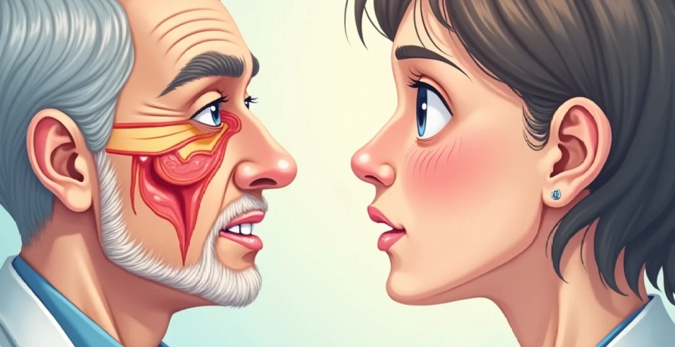
Trigeminal neuralgia and fifth cranial nerve dysfunction
Trigeminal neuralgia, whilst primarily characterised by severe facial pain, can occasionally present with accompanying numbness and dizziness. The condition typically results from vascular compression of the trigeminal nerve root, though secondary causes including multiple sclerosis, tumours, or arteriovenous malformations must be considered. When trigeminal dysfunction occurs alongside vestibular symptoms, it often suggests a more central pathological process affecting the brainstem.
The relationship between trigeminal nerve dysfunction and dizziness may seem counterintuitive, but anatomical considerations explain this association. The trigeminal nerve maintains connections with various brainstem nuclei, including those involved in vestibular processing. Additionally, the trigeminovestibular reflex pathway can be disrupted when significant trigeminal pathology is present, potentially contributing to balance disturbances.
Vestibular neuritis and eighth cranial nerve pathology
Vestibular neuritis primarily affects the vestibular portion of the eighth cranial nerve, causing severe vertigo, nausea, and imbalance. However, when facial numbness accompanies these classic vestibular symptoms, clinicians must consider more extensive pathological processes. The proximity of the facial nerve (seventh cranial nerve) to the vestibulocochlear nerve within the internal auditory canal means that inflammatory or compressive lesions can affect both structures simultaneously.
Viral infections, particularly herpes simplex virus and varicella-zoster virus, can cause simultaneous inflammation of multiple cranial nerves. This condition, known as cranial polyneuritis , can produce the combination of facial numbness, weakness, and vestibular dysfunction. The inflammatory process typically develops over days to weeks, with symptoms potentially persisting for months before resolution.
Cerebellopontine angle tumours and acoustic neuromas
Cerebellopontine angle tumours represent serious structural lesions that frequently present with progressive facial numbness and vestibular symptoms. Acoustic neuromas, the most common tumours in this region, typically begin with unilateral hearing loss and tinnitus before progressing to involve facial sensation and balance. The gradual growth of these benign tumours allows for slow adaptation, meaning symptoms may develop insidiously over months or years.
The anatomical configuration of the cerebellopontine angle creates a confined space where tumour growth inevitably affects multiple cranial nerves. As acoustic neuromas enlarge, they commonly compress the trigeminal nerve, producing facial numbness that typically begins in the distribution of the nerve’s sensory branches. Simultaneously, the expanding mass effect disrupts normal vestibular function, leading to progressive imbalance and spatial disorientation.
Large acoustic neuromas can compress the brainstem itself, potentially affecting multiple neural pathways and producing a complex constellation of neurological symptoms including facial numbness, dizziness, and coordination difficulties.
Brainstem stroke and vertebrobasilar insufficiency
Brainstem strokes represent neurological emergencies that can rapidly produce combinations of cranial nerve deficits, including facial numbness and severe dizziness. The vertebrobasilar arterial system supplies critical brainstem structures, including the trigeminal and vestibular nerve nuclei. When vascular compromise affects these regions, patients can experience sudden onset of facial sensory loss combined with vertigo, nausea, and ataxia.
Lateral medullary syndrome, also known as Wallenberg syndrome, exemplifies how brainstem vascular events can produce characteristic combinations of symptoms. This condition results from posterior inferior cerebellar artery (PICA) territory infarction, affecting the lateral medulla and producing ipsilateral facial numbness, vestibular dysfunction, and various other neurological deficits. The syndrome’s distinctive presentation includes alternating sensory loss, where facial numbness occurs on the same side as the lesion whilst body sensory loss affects the opposite side.
Vascular disorders presenting with facial paraesthesia and vertigo
Vascular pathology represents a critical category of conditions that can simultaneously affect facial sensation and vestibular function. The intricate vascular supply to the brainstem and cranial nerves makes these structures particularly vulnerable to circulatory disturbances. Understanding the vascular anatomy and pathophysiology behind these conditions is essential for recognising potentially life-threatening situations that require immediate intervention.
Posterior circulation stroke and PICA territory infarction
Posterior circulation strokes account for approximately 20% of all ischaemic strokes but carry disproportionately high morbidity and mortality rates. The vertebrobasilar system supplies critical brainstem structures, including the vestibular nuclei, trigeminal nerve nuclei, and associated pathways. When thrombotic or embolic events compromise this circulation, patients can experience rapid onset of facial numbness combined with severe vertigo and other brainstem symptoms.
PICA territory infarctions present particular diagnostic challenges because early symptoms may be mistakenly attributed to benign peripheral vestibular disorders. However, the combination of facial numbness with acute vertigo should raise immediate suspicion for brainstem pathology. Additional concerning features include dysarthria, dysphagia, ataxia, or alternating sensory patterns that distinguish central from peripheral vestibular causes.
The temporal evolution of symptoms in posterior circulation strokes can vary significantly. Some patients experience sudden, dramatic onset of neurological deficits, whilst others may have stuttering or progressive symptom development over hours or days. This variability emphasises the importance of maintaining high clinical suspicion when evaluating patients with combined facial and vestibular symptoms, particularly in those with vascular risk factors.
Vertebral artery dissection and wallenberg syndrome
Vertebral artery dissection represents a particularly serious cause of posterior circulation compromise that can produce characteristic combinations of facial numbness and vestibular dysfunction. This condition often affects younger patients without traditional vascular risk factors, making diagnosis challenging. The dissection process can occur spontaneously or following neck trauma, chiropractic manipulation, or even minor activities involving neck extension or rotation.
Wallenberg syndrome, resulting from vertebral artery dissection with subsequent PICA territory infarction, produces a distinctive clinical pattern. Patients typically experience sudden onset of severe vertigo, nausea, and vomiting, accompanied by ipsilateral facial numbness and loss of pain and temperature sensation. The combination of these symptoms with Horner’s syndrome, ataxia, and contralateral body sensory loss creates the classic presentation that neurologists recognise immediately.
Cervical artery dissection often presents with preceding neck pain or headache, which may be dismissed as benign musculoskeletal complaints. However, when these symptoms progress to include facial numbness and dizziness, immediate vascular imaging is warranted to exclude arterial dissection and guide appropriate anticoagulation therapy.
Carotid artery disease and hemodynamic compromise
Whilst carotid artery disease primarily affects anterior circulation territories, severe stenosis or occlusion can occasionally contribute to symptoms involving posterior circulation structures through various compensatory mechanisms. Hemodynamic compromise may affect the delicate vascular supply to brainstem regions, particularly when collateral circulation is inadequate or when systemic hypotension compounds the perfusion deficit.
Patients with severe bilateral carotid stenosis may experience global cerebral hypoperfusion that can manifest as dizziness, confusion, and various sensory disturbances. The facial numbness in these cases typically results from compromised perfusion to thalamic or brainstem sensory processing centres rather than direct trigeminal nerve involvement. This mechanism explains why symptoms may be bilateral or alternating rather than following classic nerve distribution patterns.
Migraine-associated vertebrobasilar insufficiency
Basilar-type migraine, previously known as basilar artery migraine, can produce dramatic combinations of brainstem symptoms including facial numbness and severe vertigo. This migraine subtype affects the vertebrobasilar circulation through vasospastic mechanisms that temporarily compromise blood flow to posterior fossa structures. The resulting symptoms can be frightening for patients and challenging for clinicians to distinguish from actual vascular events.
The pathophysiology involves cortical spreading depression that affects brainstem regions, producing transient dysfunction of multiple cranial nerve nuclei. Patients typically experience prodromal symptoms including visual disturbances, followed by the development of facial numbness, vertigo, dysarthria, and other brainstem signs. These symptoms usually resolve completely within 24 hours, distinguishing them from permanent vascular lesions.
Basilar-type migraine predominantly affects young women and can be triggered by hormonal fluctuations, stress, or specific dietary factors, making detailed history-taking crucial for accurate diagnosis.
Metabolic and systemic conditions affecting cranial nerve function
Systemic metabolic disorders can significantly impact cranial nerve function, producing combinations of facial numbness and dizziness through various pathophysiological mechanisms. These conditions often affect multiple organ systems, making comprehensive evaluation essential for accurate diagnosis and appropriate treatment. The interplay between metabolic derangements and neurological function creates complex clinical presentations that require careful analysis to identify underlying causes.
Diabetes mellitus represents the most prevalent metabolic cause of peripheral neuropathy, affecting both sensory and autonomic nervous systems. Diabetic cranial neuropathies can involve the trigeminal nerve, producing facial numbness, whilst concurrent diabetic autonomic neuropathy may contribute to orthostatic hypotension and associated dizziness. The combination of these effects creates a challenging clinical scenario where multiple pathophysiological processes contribute to symptom development.
Thyroid dysfunction, particularly hypothyroidism, can produce neurological symptoms including peripheral neuropathy and vestibular disturbances. The mechanisms involve altered protein synthesis, reduced nerve conduction velocity, and changes in inner ear fluid dynamics. Patients with severe hypothyroidism may experience myxoedema-related neuropathy affecting facial sensation whilst concurrent vestibular dysfunction contributes to balance problems and dizziness.
Vitamin deficiencies, especially B12, thiamine, and folate deficiencies, can cause peripheral neuropathies that affect cranial nerves. The trigeminal nerve’s lengthy course makes it particularly susceptible to nutritional deficiencies, whilst concurrent effects on central vestibular processing can produce dizziness and imbalance. These conditions are often reversible with appropriate supplementation, emphasising the importance of nutritional assessment in patients presenting with these symptoms.
Chronic kidney disease and uraemia can produce peripheral neuropathy through the accumulation of uremic toxins. The resulting cranial nerve dysfunction may manifest as facial numbness, whilst concurrent electrolyte imbalances and fluid retention can contribute to dizziness and balance disturbances. Dialysis patients may experience fluctuating symptoms related to fluid and electrolyte shifts during treatment sessions.
Infectious diseases causing facial numbness with vestibular symptoms
Infectious aetiologies represent important causes of concurrent facial numbness and dizziness, with viral infections being particularly common culprits. The neurotropic properties of certain viruses allow them to target specific cranial nerves or brainstem structures, producing characteristic patterns of neurological dysfunction. Understanding the temporal evolution and associated features of infectious causes helps distinguish them from other pathological processes.
Herpes zoster virus can affect multiple cranial nerves simultaneously, producing Ramsay Hunt syndrome when the facial and vestibulocochlear nerves are involved. This condition presents with characteristic vesicular eruptions in the external auditory canal, accompanied by facial weakness, sensory loss, hearing impairment, and vestibular dysfunction. The combination of facial numbness with severe vertigo in the setting of characteristic skin lesions makes diagnosis relatively straightforward.
Lyme disease, caused by Borrelia burgdorferi infection, can produce neurological complications including cranial neuropathies and central nervous system involvement. Neuroborreliosis may present with facial numbness due to trigeminal nerve involvement, whilst concurrent inflammation affecting vestibular pathways produces dizziness and balance disturbances. The diagnosis requires consideration of tick exposure history and appropriate serological testing.
Epstein-Barr virus and cytomegalovirus infections can cause inflammatory conditions affecting multiple cranial nerves. These viral infections may produce post-infectious inflammatory syndromes that target peripheral nerves or brainstem structures. Patients typically present with preceding febrile illness followed by the development of neurological symptoms including facial numbness and vestibular dysfunction.
Meningitis and encephalitis represent serious infectious conditions that can affect cranial nerve function through direct inflammatory involvement or increased intracranial pressure effects. Bacterial, viral, or fungal pathogens can produce meningeal inflammation that extends to cranial nerve roots, causing dysfunction of both sensory and vestibular pathways. The combination of facial numbness and dizziness with headache, fever, and altered mental status suggests these serious infectious processes.
Medication-induced peripheral neuropathy and ototoxicity
Pharmaceutical agents represent significant iatrogenic causes of both peripheral neuropathy affecting facial sensation and ototoxicity producing vestibular dysfunction. The concurrent development of these symptoms following medication initiation or dose escalation provides important diagnostic clues. Understanding the mechanisms by which various drugs affect cranial nerve function enables clinicians to identify reversible causes and implement appropriate management strategies.
Chemotherapeutic agents, particularly platinum-based compounds like cisplatin and carboplatin, are well-recognised causes of both peripheral neuropathy and ototoxicity. These medications produce dose-dependent toxicity that can affect the trigeminal nerve, causing facial numbness, whilst simultaneously damaging inner ear structures responsible for hearing and balance. The resulting combination of facial sensory loss and vestibular dysfunction can significantly impact quality of life for cancer patients.
Aminoglycoside antibiotics represent another important class of medications that can produce concurrent cranial nerve toxicity. These agents preferentially accumulate in inner ear tissues, causing damage to both cochlear and vestibular hair cells. When high doses or prolonged treatment courses are employed, additional effects on peripheral nerves may occur, producing facial numbness alongside the characteristic vestib
ular dysfunction.
Antiepileptic drugs, including phenytoin, carbamazepine, and newer agents like lamotrigine, can cause peripheral neuropathy as a dose-related side effect. The mechanism involves interference with sodium channel function and altered nerve membrane stability. When these medications affect the trigeminal nerve, patients may experience facial numbness, whilst concurrent central nervous system effects can contribute to dizziness and balance disturbances through altered cerebellar function.
Loop diuretics, particularly furosemide and bumetanide, can produce ototoxicity when used in high doses or in patients with renal impairment. The resulting damage to inner ear structures produces both hearing loss and vestibular dysfunction, manifesting as dizziness and imbalance. When combined with electrolyte disturbances commonly associated with diuretic use, patients may experience additional neurological symptoms including peripheral neuropathy affecting facial sensation.
The temporal relationship between medication initiation and symptom onset provides crucial diagnostic information, with most drug-induced neuropathies developing weeks to months after treatment commencement.
Antiretroviral medications used in HIV treatment can cause peripheral neuropathy through mitochondrial toxicity mechanisms. Nucleoside reverse transcriptase inhibitors, particularly stavudine and didanosine, are associated with significant neuropathic complications that can affect cranial nerves. The resulting facial numbness, combined with central nervous system effects of HIV infection itself, creates complex clinical presentations requiring careful evaluation to distinguish drug-induced effects from disease progression.
Diagnostic approaches for combined facial and vestibular dysfunction
The diagnostic evaluation of patients presenting with concurrent facial numbness and dizziness requires a systematic approach that considers the broad differential diagnosis whilst prioritising potentially life-threatening conditions. The initial assessment must rapidly identify emergency situations requiring immediate intervention, particularly brainstem strokes or other vascular events that can cause irreversible neurological damage if treatment is delayed.
Clinical history-taking forms the cornerstone of diagnostic evaluation, with particular attention to symptom onset, progression, and associated features. Sudden onset of symptoms suggests vascular aetiologies, whilst gradual progression over weeks to months may indicate tumours or degenerative conditions. The presence of additional symptoms such as headache, visual disturbances, or weakness provides valuable localising information that guides subsequent investigations.
Physical examination must include comprehensive neurological assessment focusing on cranial nerve function, coordination, and balance testing. The pattern of facial sensory loss helps distinguish between peripheral trigeminal nerve dysfunction and central brainstem pathology. Vestibular examination techniques, including the head impulse test and positional manoeuvres, can differentiate peripheral from central causes of dizziness and guide further evaluation.
Neuroimaging represents a critical component of the diagnostic workup, with MRI being the preferred modality for evaluating posterior fossa structures and brainstem pathology. T2-weighted and FLAIR sequences are particularly sensitive for detecting demyelinating lesions, whilst diffusion-weighted imaging can identify acute ischaemic changes within minutes of symptom onset. Gadolinium-enhanced sequences help identify inflammatory conditions, tumours, or vascular abnormalities affecting cranial nerves.
Audiometry and vestibular function testing provide objective measures of eighth cranial nerve function and can help localise pathology within the auditory and vestibular systems. Pure tone audiometry, tympanometry, and acoustic reflex testing evaluate different components of hearing function, whilst electronystagmography or videonystagmography can quantify vestibular dysfunction and distinguish peripheral from central causes.
Laboratory investigations should be tailored to the clinical presentation but may include inflammatory markers, autoimmune panels, vitamin levels, and infectious disease screening. Cerebrospinal fluid analysis may be necessary when infectious or inflammatory conditions are suspected, providing information about cell counts, protein levels, and pathogen-specific markers. Genetic testing may be considered in patients with family histories of inherited neuropathies or degenerative conditions.
Electrophysiological studies, including nerve conduction studies and electromyography, can evaluate peripheral nerve function and help distinguish between axonal and demyelinating neuropathies. When facial weakness accompanies numbness, these studies can localise lesions to specific nerve segments and provide prognostic information about recovery potential. Brainstem auditory evoked potentials may be useful for evaluating eighth cranial nerve function in patients with hearing loss or vestibular symptoms.
The integration of clinical findings with diagnostic test results requires careful consideration of the entire clinical picture. False-positive and false-negative results can occur with any diagnostic modality, making clinical correlation essential for accurate diagnosis. When initial investigations are normal but clinical suspicion remains high for serious pathology, repeat imaging or additional testing may be necessary to exclude evolving conditions or identify subtle abnormalities not apparent on initial studies.