Nail injuries represent one of the most common traumatic conditions affecting the hands and feet, accounting for approximately 5% of all emergency department visits. Whether caused by crushing trauma, avulsion injuries, or chemical exposure, damage to the nail unit raises immediate questions about regeneration potential and long-term aesthetic outcomes. The human nail apparatus demonstrates remarkable regenerative capacity, yet the extent of recovery depends heavily on the specific structures affected and the severity of the initial trauma.
Understanding nail regrowth after injury requires comprehensive knowledge of the complex anatomical structures involved and the intricate biological processes that govern nail formation. Modern medical approaches have evolved significantly, offering both surgical and conservative treatment options that can dramatically influence regenerative outcomes. The prognosis for nail regrowth varies considerably based on injury classification, patient factors, and the timeliness of appropriate intervention.
Nail anatomy and regeneration physiology after traumatic injury
The nail unit comprises several interconnected structures that work in harmony to produce the protective nail plate we observe. Successful nail regeneration depends fundamentally on the integrity of these anatomical components, particularly the nail matrix, which serves as the primary site of nail cell production. When trauma occurs, the extent of damage to each structure determines the regenerative potential and ultimate aesthetic outcome.
Nail matrix structure and keratinocyte production mechanisms
The nail matrix functions as the powerhouse of nail production , containing specialised keratinocyte stem cells that undergo continuous division and differentiation. This structure extends from the proximal nail fold to approximately 2-3 millimetres beyond the visible lunula, creating what medical professionals term the “critical zone” for nail regeneration. The germinal matrix, located in the most proximal portion, contributes approximately 90% of the nail plate thickness through rapid cell division and subsequent keratinisation processes.
During the regeneration process, matrix keratinocytes follow a precisely orchestrated programme of proliferation, differentiation, and migration. These cells produce specialised proteins including hard keratins and nail-specific structural proteins that provide the characteristic strength and flexibility of healthy nails. Following trauma, the matrix cells must re-establish their normal proliferative patterns, which typically requires 4-6 weeks before visible nail growth becomes apparent.
Eponychium and hyponychium healing response patterns
The eponychium and hyponychium serve as critical barriers protecting the nail unit from environmental pathogens and mechanical trauma. These structures demonstrate remarkable adaptive responses following injury, often developing temporary hyperkeratotic changes that provide additional protection during the vulnerable healing phase. The eponychium, commonly referred to as the cuticle, plays a particularly important role in maintaining the seal between the nail plate and surrounding soft tissues.
Research indicates that proper healing of these structures significantly influences long-term nail quality and appearance. Disruption of the eponychium can lead to chronic bacterial colonisation and subsequent nail deformities, whilst hyponychium damage may result in painful hypersensitivity and increased susceptibility to subungual haemorrhages. Clinical studies demonstrate that careful attention to these anatomical regions during treatment can improve regenerative outcomes by up to 40%.
Vascular supply to the nail unit during recovery
The nail unit receives its blood supply through an intricate network of vessels originating from the digital arteries. This vascular architecture demonstrates remarkable redundancy, with multiple anastomotic connections ensuring continued perfusion even after significant trauma. The rich capillary plexus beneath the nail matrix proves particularly crucial for regeneration, as these vessels deliver essential nutrients and growth factors required for keratinocyte proliferation.
Following injury, the vascular response typically progresses through distinct phases of inflammation, proliferation, and remodelling. Initial vasoconstriction helps limit bleeding, followed by vasodilation and increased permeability that facilitates inflammatory cell recruitment. New vessel formation, or angiogenesis, becomes apparent within 48-72 hours post-injury, with peak vascularisation occurring during the second week of healing. This enhanced blood supply can persist for several months, contributing to the characteristic pink or red appearance often observed in regenerating nails.
Neural innervation impact on nail regrowth potential
Sensory innervation of the nail unit derives primarily from branches of the median, ulnar, and radial nerves in the fingers, with corresponding innervation patterns in the toes. These neural pathways not only provide sensation but also contribute to the regulation of local blood flow through neurogenic mechanisms. Denervation following trauma can significantly impact regenerative capacity, as neural influences play important roles in growth factor release and cellular metabolism.
Studies examining nail regeneration in denervated digits reveal consistently slower growth rates and altered nail plate characteristics. The absence of normal neural input appears to affect matrix cell proliferation patterns and may contribute to the development of dystrophic changes in the regenerated nail. However, the nail unit demonstrates considerable adaptability, and partial reinnervation often occurs spontaneously over time, leading to gradual improvement in nail quality and growth rates.
Classification of nail injuries and regeneration prognosis
Medical professionals utilise comprehensive classification systems to categorise nail injuries based on anatomical involvement, mechanism of trauma, and severity of tissue damage. This systematic approach enables accurate prognosis determination and guides treatment decision-making. The most widely accepted classification system divides nail injuries into four primary categories: avulsion injuries, crush injuries, lacerations, and chemical or thermal burns. Each category demonstrates distinct regenerative patterns and requires specific therapeutic approaches.
Avulsion injuries: complete vs partial nail plate detachment
Avulsion injuries occur when mechanical forces separate the nail plate from its underlying attachments, either partially or completely. Complete avulsions involve total separation of the nail plate from both the nail bed and matrix, whilst partial avulsions maintain some degree of attachment. These injuries commonly result from catching fingernails in doors, machinery accidents, or sports-related trauma.
The regenerative prognosis for avulsion injuries depends heavily on the extent of matrix involvement and the condition of the remaining nail bed. Complete avulsions with intact nail beds typically demonstrate excellent regenerative potential, with new nail growth becoming visible within 6-8 weeks. However, injuries involving significant matrix damage may result in permanent nail deformities or complete failure of regeneration. Recent studies indicate that approximately 85% of complete avulsions with preserved matrix tissue achieve satisfactory cosmetic outcomes within 6-12 months.
Complete nail avulsion with intact nail matrix demonstrates regeneration success rates exceeding 85% when appropriate treatment protocols are implemented within the first 24 hours post-injury.
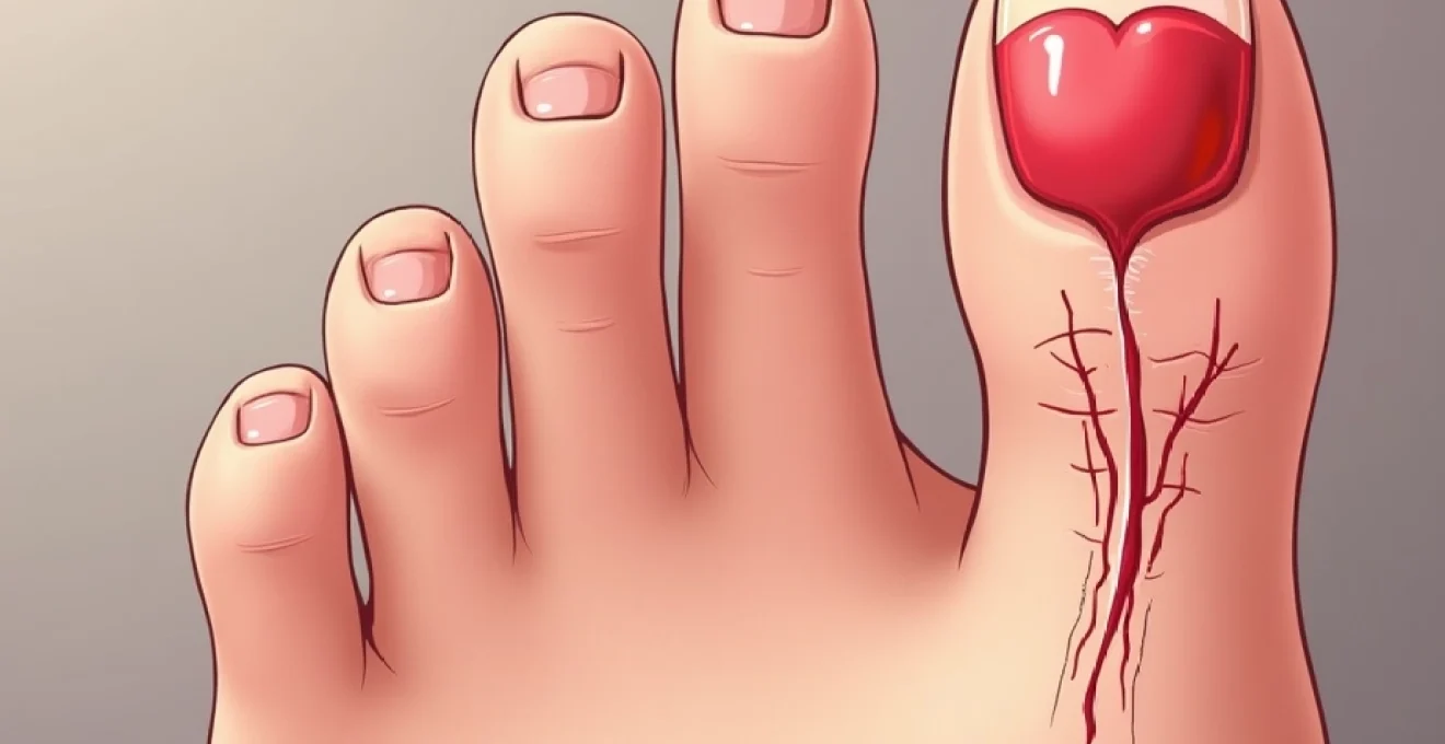
Crush injuries and subungual haematoma formation
Crush injuries represent the most common mechanism of nail trauma, frequently resulting from heavy objects falling onto digits or fingers being caught in closing doors. These injuries typically produce subungual haematomas, characterised by blood accumulation beneath the nail plate. The pressure created by trapped blood can cause significant pain and may compromise local circulation if left untreated.
The presence of subungual haematoma covering more than 50% of the nail surface often indicates underlying nail bed laceration or matrix injury. Prompt decompression through trephination can provide immediate pain relief and may prevent secondary complications such as nail plate separation or infection. Regeneration following crush injuries varies considerably, with minor injuries showing complete recovery whilst severe crushing may result in permanent nail dystrophy or loss.
Laceration patterns affecting nail bed integrity
Sharp trauma resulting in nail bed lacerations requires careful assessment to determine the extent of tissue damage and the optimal repair strategy. Simple lacerations confined to the sterile matrix typically heal well with conservative management, whilst complex injuries involving the germinal matrix often require surgical intervention to achieve optimal outcomes.
Stellate lacerations, characterised by multiple tear patterns radiating from a central point, present particular challenges for reconstruction. These injuries often result from glass cuts or machinery accidents and may involve loss of nail bed tissue. The regenerative potential depends on the surgeon’s ability to restore anatomical relationships and maintain adequate blood supply to the repaired tissues. Clinical data suggests that precise surgical repair of nail bed lacerations can achieve normal nail regrowth in approximately 75-90% of cases.
Chemical burns and thermal damage to nail matrix
Chemical and thermal injuries to the nail unit often produce devastating damage to the matrix and surrounding tissues. Strong acids, alkalis, or extreme temperatures can cause coagulative necrosis of the nail-forming tissues, resulting in permanent loss of regenerative capacity. These injuries frequently occur in industrial settings or as a result of domestic accidents involving cleaning products or hot appliances.
The depth of tissue damage determines regenerative potential, with superficial injuries showing good recovery whilst full-thickness burns typically result in scarring and nail dystrophy. Chemical burns may continue to cause tissue damage for hours after initial exposure, making prompt irrigation and neutralisation crucial for limiting the extent of injury. Studies indicate that chemical burns affecting more than 75% of the matrix area rarely achieve complete nail regeneration, though partial recovery may still occur.
Clinical assessment protocols for nail regeneration evaluation
Comprehensive clinical assessment forms the foundation of successful nail injury management and accurate prognosis determination. The evaluation process must systematically examine all components of the nail unit whilst considering patient factors that may influence healing capacity. Modern assessment protocols incorporate both clinical examination techniques and advanced imaging modalities to provide detailed information about injury extent and tissue viability.
Initial assessment begins with careful history taking to establish the mechanism of injury, time elapsed since trauma, and any previous nail disorders or systemic conditions that might affect healing. Physical examination requires adequate lighting and magnification to visualise fine anatomical details. The clinician must assess nail plate integrity, examine the nail bed for lacerations or haematomas, and evaluate matrix function through inspection of the lunula and proximal nail fold.
Systematic documentation of injury patterns using standardised terminology ensures consistent communication between healthcare providers and facilitates outcome comparison across different treatment modalities. Digital photography has become an invaluable tool for documenting initial injury appearance and tracking regenerative progress over time. High-resolution images allow detailed analysis of nail growth patterns and help identify subtle deformities that might not be apparent during routine clinical examination.
Advanced imaging techniques, including high-frequency ultrasound and optical coherence tomography, are increasingly utilised to assess nail bed integrity and matrix viability in complex cases. These non-invasive modalities can identify subtle tissue damage not apparent on clinical examination and may help guide surgical decision-making. Research studies have demonstrated that ultrasound assessment of nail bed thickness and vascularity correlates well with regenerative outcomes.
Pain assessment requires careful attention, as the intensity and character of discomfort can provide valuable diagnostic information. Sharp, localised pain often indicates active tissue damage or infection, whilst diffuse throbbing may suggest haematoma formation or compromised circulation. Functional assessment examines grip strength, pinch patterns, and fine motor control, as nail injuries can significantly impact hand function even after successful regeneration.
Surgical interventions and nail bed reconstruction techniques
Surgical management of nail injuries has evolved significantly over recent decades, with refined techniques enabling improved functional and aesthetic outcomes. The decision to pursue surgical intervention depends on multiple factors including injury severity, anatomical structures involved, patient age and occupation, and expected functional demands. Modern surgical approaches emphasise preservation of viable tissue, anatomical restoration, and optimisation of regenerative potential.
Primary nail bed repair using absorbable sutures
Primary repair of nail bed lacerations represents the gold standard for achieving optimal regenerative outcomes. The technique involves meticulous debridement of non-viable tissue followed by precise approximation of wound edges using fine absorbable sutures. Surgeons typically employ 6-0 or 7-0 chromic gut or polyglycolic acid sutures, which provide adequate tensile strength whilst avoiding the need for subsequent removal.
The surgical approach requires complete nail plate removal to provide adequate visualisation of the injury. Following thorough irrigation and debridement, the nail bed is carefully inspected for retained foreign material or devitalised tissue. Precise suture placement ensures anatomical alignment of the nail bed ridges and grooves, which is crucial for normal nail plate formation. Studies demonstrate that meticulous primary repair can achieve normal nail regrowth in over 90% of simple lacerations.
Post-operative management involves careful wound protection and monitoring for signs of infection or healing complications. The replaced nail plate serves as a biological dressing, protecting the repaired nail bed whilst providing a template for new nail growth. Regular follow-up examinations allow early detection of healing problems and enable timely intervention if complications arise.
Split-thickness skin grafting for extensive nail bed loss
Cases involving significant nail bed tissue loss require reconstructive techniques to restore the anatomical foundation necessary for nail regeneration. Split-thickness skin grafting has emerged as a reliable method for replacing missing nail bed tissue, with grafts typically harvested from the hypothenar eminence or plantar foot surface. These donor sites provide skin with similar thickness and texture characteristics to the native nail bed.
The grafting procedure requires careful preparation of the recipient bed to ensure optimal graft survival. All non-viable tissue must be debrided, and the wound bed should demonstrate adequate vascularity to support graft integration. Precise graft sizing and positioning are crucial for achieving satisfactory outcomes, as oversized grafts may buckle whilst undersized grafts leave defects that heal by secondary intention.
Graft survival rates typically exceed 85% when proper technique is employed and post-operative care protocols are followed. However, nail regeneration over grafted areas often demonstrates altered characteristics including increased thickness, irregular surface texture, or abnormal pigmentation. Despite these limitations, split-thickness grafting can restore protective function and provide acceptable aesthetic outcomes in cases where primary repair is not feasible.
Composite grafting techniques from toe to fingernail units
Complex fingernail injuries involving extensive tissue loss may benefit from composite grafting techniques using donor tissue from the toes. This approach involves transfer of intact nail bed, matrix, and nail plate from a toe donor site to reconstruct the damaged fingernail unit. The technique is particularly valuable for high-demand patients such as musicians or surgeons where optimal fingernail function is crucial.
Toe-to-finger nail transplantation requires sophisticated microsurgical techniques to achieve vascular anastomoses and ensure graft survival. The second toe typically serves as the optimal donor site due to size compatibility and minimal functional impact. Successful composite grafting can achieve remarkable functional and aesthetic outcomes, with transplanted nails demonstrating normal growth patterns and appearance.
Long-term studies of composite nail grafting report success rates approaching 80% for properly selected cases. However, the procedure requires significant surgical expertise and carries risks associated with microsurgical techniques. Patient selection criteria include adequate vascular status, realistic expectations, and willingness to accept potential donor site morbidity.
Microsurgical approaches for nail matrix restoration
Severe matrix injuries may require microsurgical reconstruction to restore nail-forming capacity. These technically demanding procedures involve transfer of vascularised tissue containing viable nail matrix cells from uninjured digits or toes. The approach is reserved for cases where conventional repair techniques are unlikely to achieve satisfactory outcomes.
Microsurgical matrix reconstruction requires specialised equipment and extensive surgical experience. The procedure involves harvesting a composite tissue graft containing matrix tissue, nail bed, and associated vascular supply. Precise vascular anastomosis ensures graft survival and maintains the cellular environment necessary for nail formation. Success rates vary considerably based on surgeon experience and case complexity, with reported success rates ranging from 60-85%.
Microsurgical nail matrix reconstruction represents the most advanced technique available for restoring nail-forming capacity in severely damaged digits, achieving success rates of 60-85% in specialised centres.
Non-surgical management and conservative treatment protocols
Conservative management remains the preferred approach for many nail injuries, particularly those with intact nail matrix and minimal tissue loss. Non-surgical protocols focus on optimising the natural healing environment, preventing complications, and supporting the regenerative process through appropriate wound care and patient education. The success of conservative treatment depends heavily on accurate injury assessment and patient compliance with prescribed care regimens.
Initial wound management emphasises thorough cleaning and debridement of foreign material whilst preserving viable tissue. Gentle irrigation with saline solution removes debris without causing additional trauma to delicate regenerating structures. Antimicrobial prophylaxis may be indicated for contaminated wounds or patients with increased infection risk, though routine antibiotic use is not recommended for clean injuries.
Protective dressing techniques play a crucial role in conservative management success. Modern wound dressings incorporate advanced materials that maintain optimal moisture levels whilst providing barrier protection against bacterial contamination. Hydrocolloid and foam dressings have demonstrated superior outcomes compared to traditional gauze
dressings, promoting faster healing and reducing infection rates by maintaining appropriate moisture balance and temperature regulation.
Patient education forms a cornerstone of conservative management success. Individuals must understand the importance of keeping the injured area clean and dry, recognising signs of infection, and adhering to prescribed activity restrictions. Regular monitoring protocols enable early detection of healing complications, with patients instructed to observe for increased pain, swelling, discharge, or changes in nail bed colour. Studies indicate that comprehensive patient education can reduce complication rates by up to 30% compared to standard care approaches.
Activity modification during the healing phase helps prevent re-injury and optimises regenerative conditions. Patients should avoid activities that could traumatise the healing nail unit, including contact sports, heavy manual labour, or prolonged water exposure. Protective splinting may be beneficial for certain injury patterns, particularly those involving the fingertips where accidental trauma is more likely to occur.
Complications and abnormal nail regrowth patterns
Despite optimal treatment approaches, nail injuries can result in various complications that affect both function and appearance. Understanding these potential complications enables clinicians to counsel patients appropriately and implement preventive strategies where possible. The likelihood of complications correlates directly with injury severity, patient age, underlying health conditions, and adherence to treatment protocols.
Pterygium formation and scar tissue development
Pterygium formation represents one of the most common complications following nail bed injuries, characterised by abnormal adhesion between the eponychium and nail bed. This condition occurs when healing tissues form aberrant connections, creating a web-like structure that advances across the nail surface. Pterygium development typically becomes apparent 4-8 weeks after injury and may continue to progress if left untreated.
The pathophysiology involves disruption of the normal barrier between the nail fold and nail bed, allowing inflammatory cells and fibroblasts to bridge the anatomical divide. As these tissues mature and contract, they create the characteristic adherent band that restricts normal nail growth. Minor pterygia may cause cosmetic concerns but limited functional impairment, whilst extensive formations can completely prevent nail regeneration.
Prevention strategies focus on maintaining proper anatomical relationships during the healing phase. Careful wound management and appropriate use of barrier dressings can help prevent abnormal tissue adhesions. When pterygium formation does occur, treatment options range from conservative management with topical therapies to surgical excision and reconstruction. Studies demonstrate that early intervention within the first 3 months of pterygium development achieves better outcomes than delayed treatment approaches.
Split nail deformities and longitudinal ridging
Split nail deformities occur when trauma damages the nail matrix in a linear pattern, resulting in a permanent groove or complete division of the nail plate. These deformities typically develop following sharp injuries that create focal areas of matrix scarring or destruction. The resulting nail demonstrates a characteristic longitudinal split that may extend from the cuticle to the free edge, creating functional and aesthetic concerns for patients.
Longitudinal ridging represents a related but distinct phenomenon characterised by parallel grooves running the length of the nail plate. These ridges result from irregular matrix function following injury, creating variations in nail plate thickness and surface texture. Mild ridging may be barely perceptible and cause minimal concern, whilst severe cases can create deep furrows that are prone to catching on clothing or splitting further.
The severity of split nail deformities depends on the width and depth of matrix damage. Narrow splits affecting less than 2mm of matrix width may show gradual improvement over time as surrounding tissues compensate for the damaged area. However, wider defects typically result in permanent deformities that may require surgical correction if functional impairment is significant. Recent advances in matrix reconstruction techniques have shown promising results for correcting established split nail deformities.
Longitudinal nail ridging affects approximately 15-20% of patients following significant nail matrix trauma, with severity correlating directly to the extent of initial tissue damage and adequacy of primary repair.
Onychogryphosis and nail plate thickening disorders
Onychogryphosis, commonly referred to as “ram’s horn nail,” represents a severe form of nail dystrophy characterised by massive thickening and curved overgrowth of the nail plate. This condition most frequently affects toenails and may develop months to years after significant trauma to the nail matrix. The pathophysiology involves disrupted matrix function leading to abnormal keratin production and altered nail plate architecture.
The development of onychogryphosis typically follows a predictable pattern beginning with mild nail thickening and progressing to severe deformity if left untreated. Early signs include increased nail plate thickness, yellowing discoloration, and slight curvature. As the condition advances, the nail develops a characteristic horn-like appearance with extreme thickness and spiral curvature that can impair footwear fitting and cause significant discomfort.
Contributing factors to onychogryphosis development include repeated microtrauma from ill-fitting shoes, fungal infections, and systemic conditions such as psoriasis or peripheral vascular disease. Age represents a significant risk factor, with the condition predominantly affecting elderly patients who may have reduced ability to maintain proper nail care. The combination of trauma-induced matrix damage and these predisposing factors creates an environment conducive to abnormal nail growth patterns.
Management of onychogryphosis requires a multifaceted approach addressing both the mechanical and underlying biological factors. Regular professional nail trimming using specialised equipment can maintain manageable nail length and prevent complications such as ingrown nails or pressure ulcers. In severe cases, partial or complete nail removal may be necessary, with chemical or surgical matrix destruction preventing recurrence. Studies indicate that early intervention can prevent progression to severe deformity in approximately 70% of cases.
Prevention strategies emphasise proper nail care, appropriate footwear selection, and prompt treatment of predisposing conditions. Patient education regarding nail trimming techniques and recognition of early warning signs enables timely intervention before irreversible changes occur. For individuals at high risk, regular podiatric care can identify developing problems and implement preventive measures to maintain nail health and function.